-
Transparencies

Two lily blossoms © Julian Köpke -
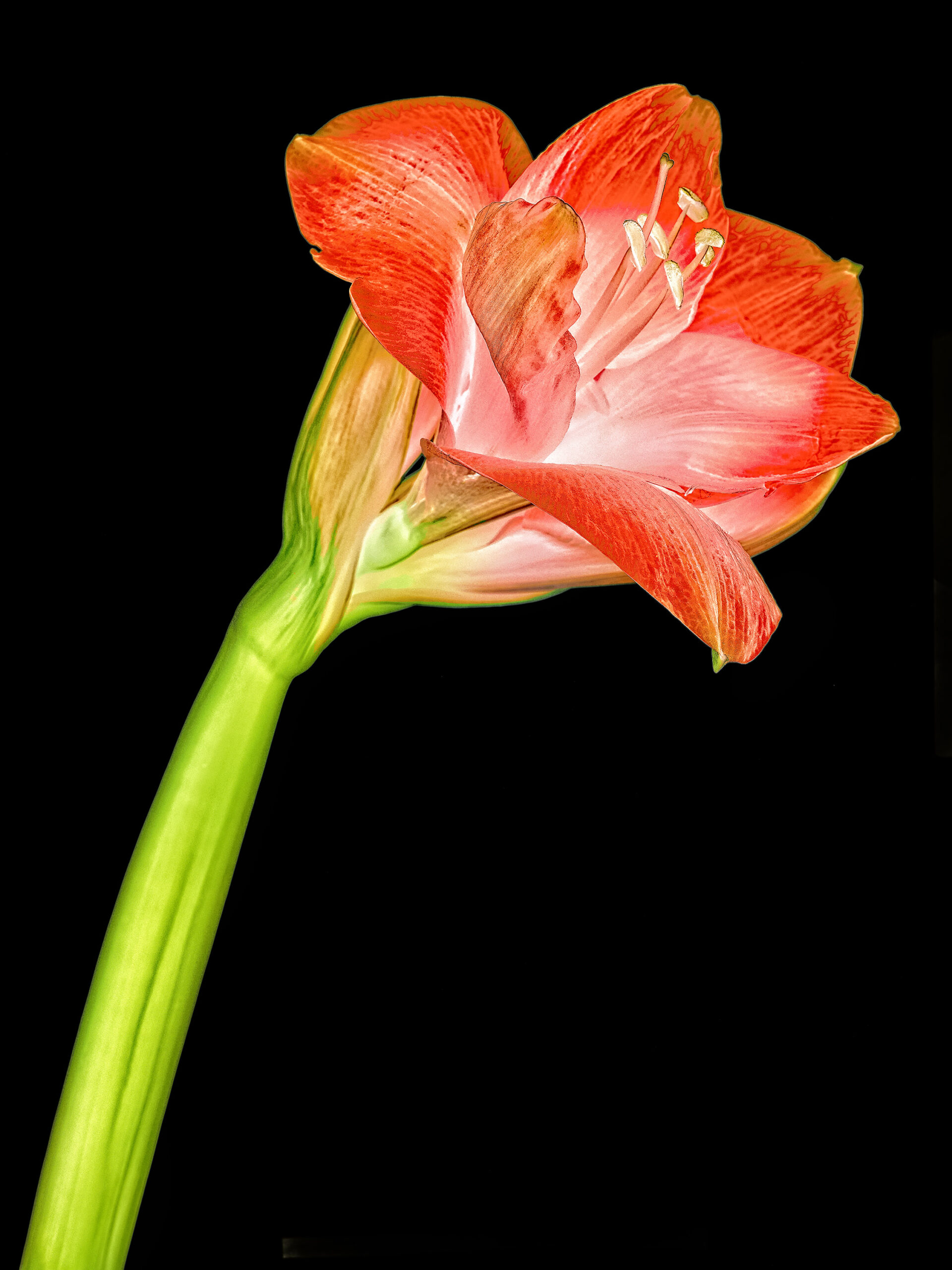
Amaryllis

Amaryllis Lab color inversion photography © Julian Köpke 
Amaryllis X-ray mammography photogram © Julian Köpke 
Amaryllis X-ray mammography fusion photography texturized © Julian Köpke -
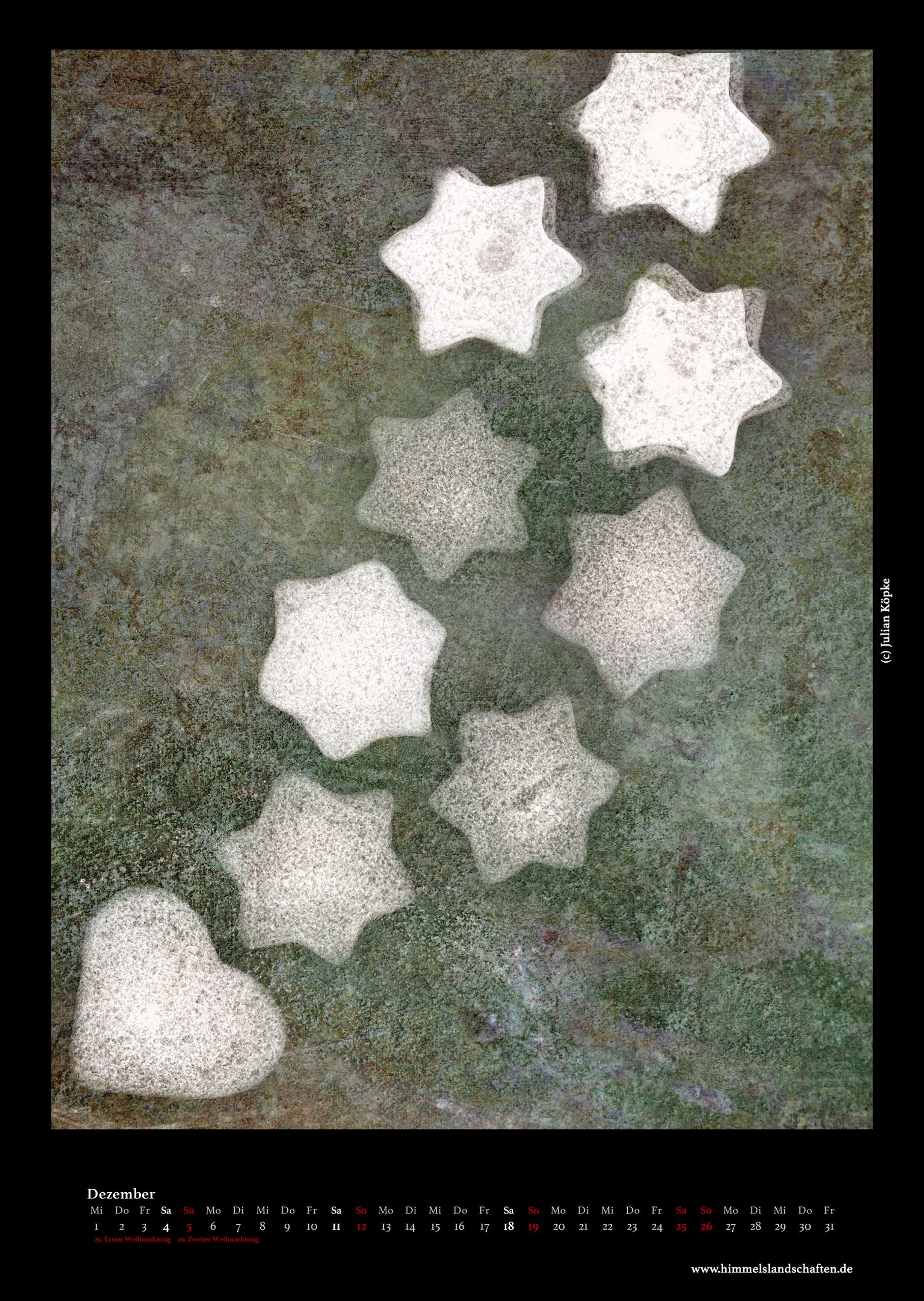
Calendar 2021
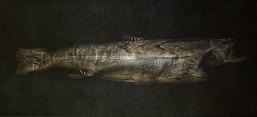
Trout X-ray fusion photo © Julian Köpke 
Black swan: pumpkins and fir cones X-ray photo © Julian Köpke 
-
Oak leaves with acorns

Oak leaves with acorns X-ray photo © Julian Köpke 
Oak leaves with acorns X-ray photo L-inversion © Julian Köpke 
Oak leaves with acorns X-ray fusion photo © Julian Köpke -
New X-ray fusion photo compositions

Fusion X-ray photo of bananas © Julian Köpke 
Fruit bowl X-ray fusion photo © Julian Köpke 
X-ray fusion photo of lychees in a wooden bowl © Julian Köpke 
X-ray fusion photo of lychees and fruit in a wooden bowl © Julian Köpke 
Fruit X-ray fusion photo © Julian Köpke -
Snail shells X-ray fusion photos

X-ray fusion photo snail shells composition II © Julian Köpke 
X-ray fusion photo snail shells composition III © Julian Köpke 
X-ray fusion photo snail shells composition IV © Julian Köpke -
X-ray fusion photo of a sphere of snail shells

X-ray fusion photo of a sphere of snail shells © Julian Köpke 
Sphere of snail shells © Julian Köpke -
Calendar 2020

-
New wine X-ray photo of Pinot Noir

Pinot noir X-ray fusion image © Julian Köpke -
Spider conch X-ray fusion photo

Fusion image spider conch X-ray and photo seen from the bottom © Julian Köpke 
Fusion image spider conch (lambis lambis) X-ray and photo seen from above © Julian Köpke 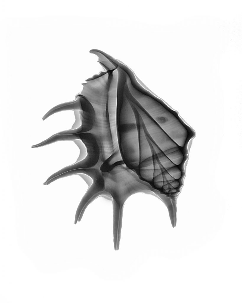
Spider conch (lambis lambis) © Julian Köpke 
Spider conch (lambis lambis) © Julian Köpke


