• flowers,  Fusion imaging,  Lightbox,  X-Ray

    Orchid X-ray fusion photo

    It feels like very long ago. Harold and I were taking the shots and X-rays of new compositions last week of  April this year. Our first try was an orchid with two stems. The transparency effect is very much augmented using an X-ray. A stem behind petals doesn’t show easily in HDR light box photography.

    With a Phase One camera at my disposal a strong crop of the composition shows the tenderness of our orchid much better. With a resolution still sufficient.

    Orchid fusion X-ray photo © Julian Köpke
  • Fusion imaging,  Lightbox,  X-Ray

    Purple Clematis

    Clematis is a reliably blossoming flower in our garden. Every year we look forward to her blooms for many weeks. Photographing flowers means sacrificing beautiful little things. It took me some time to go there.

    With growing experience I feel less pain to sacrifice a bloom for artistic purposes. It relieves me a little, that I have the blooms swum after my photo and X-ray sessions in a soup-plate filled with water which is in the kitchen. Many people like the floating blooms in a soup-plate, if they are in a break.

    The HDR series of my composition with three clematis gave me a hard time. Although a tripod is indispensable and always used, a small pixel shift between exposures was perceivable. After fixing this, light, color and structure was processed for an HDR image.

    The X-ray of the three clematis was performed as mammography due to the size of my composition. The fusion image can be understood as a texturized HDR by means of a radiograph. But there is no unique solution to all compositions. The best solution has to be found out individually.

    After all, the clematis look as light as a feather in this image. It was worth it.

    Three purple Clematis fusion X-ray photo © Julian Köpke
  • flowers,  Fusion imaging

    Purple Clematis

    Long lasting blossoms, turning up every year: my purple clematis in our garden.

    It was my third X-ray session with flowers this week. Third fusion imaging attempt. After blue cornflower and blue aquilegia now a purple clematis. Big data on my hard disk.

    Today we did it with mammography at 30 kV and 50 mAs. Lower noise ! Here is the positive representation of a single clematis:

    Clematis X-ray photo © Julian Köpke

    I processed the lightbox HighKey series with a mask. There was a shift of 2 or 3 pixels from the lightest to the darker images. So I processed everything a second time to compensate for the shift. The HDR image shows a cut stalk. Photoshop is made for this.

    Purple Clematis © Julian Köpke

    The stalk can be lengthened like in the preceding X-ray. The fusion image shows hidden leaves, the core of the blossom and stalks much better:

    Purple Clematis X-ray fusion photo © Julian Köpke

    The flower looks pretty fragile now, close to its natural appearance.

  • Fusion imaging,  Lightbox,  X-Ray

    Spring and X-ray fusion photos

    First flowers in spring show up. With much support from my colleagues I’m able to do some fusion images. We all would like to have another calendar.

    Preparing the lightbox, the X-ray machines, my camera and picking out the data is a bunch of hassle.

    My personal favorite is the blue cornflower. It looks like a print of an old botanic book:

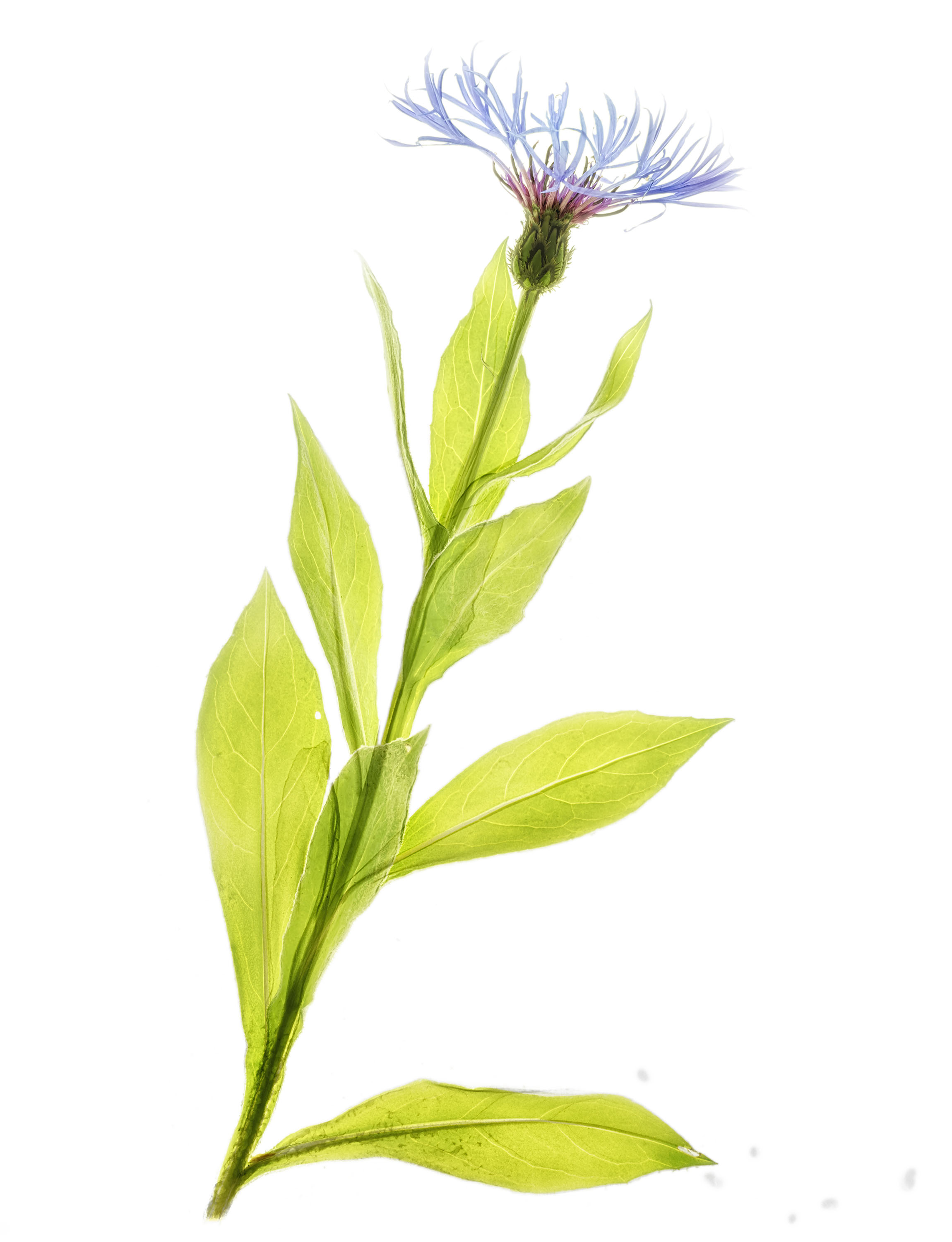
    Cornflower X-ray fusion photo © Julian Köpke

    The next day I turned my attention to our white and blue Aquilegias. No chance to process the raw data yesterday. Eventually, there was a chance today, after quite a bit of tedious work at my desk:

    Blue aquilegia X-ray fusion photo © Julian Köpke