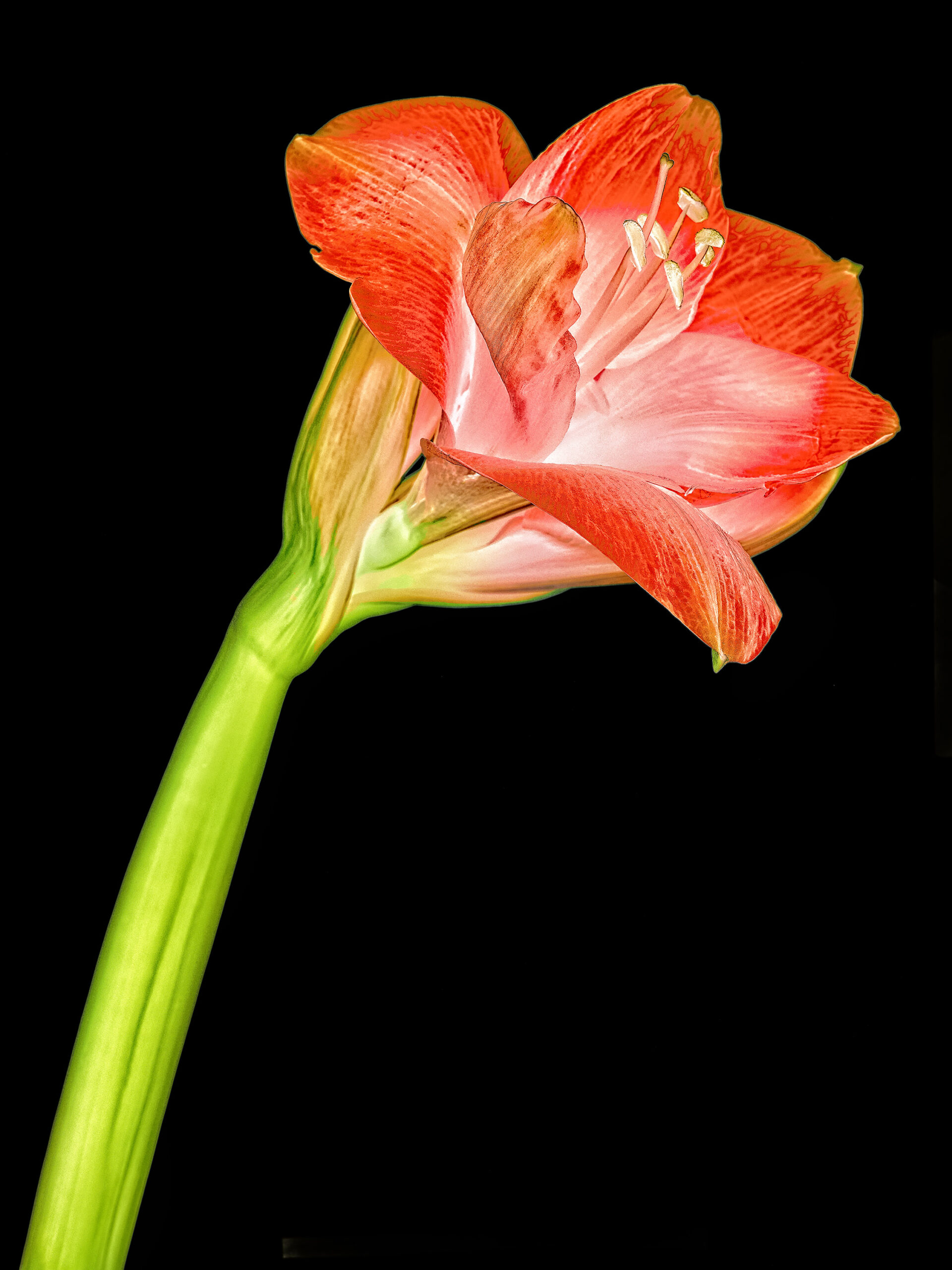
flowers,  Fusion imaging,  Lightbox,  Macro,  Texture

Amaryllis

The format for recording an image can be very variable when fusion imaging is involved. In the example below, the image on the left is taken with a Phase One IQ4. The sensor size is 44mm x 55mm. The X-ray image on the right was taken with a Hologic mammography system with a detector size of 24cm x 30cm. The aspect ratio of both images is 4:5. The pixel size of the IQ4 is 6.5µm, the pixel of the mammography is 70µm long. Nevertheless, the images can be fused well.

Amaryllis Lab color inversion photography © Julian Köpke
Amaryllis X-ray mammography photogram © Julian Köpke

I added some texture to the fusion image because it made it more appealing. Due to the fusion of the image from „visible light“ and the image with „X-ray light“, the definition of whether it is a medium format image or a large format image is no longer meaningfully applicable. It is simply an image.

Amaryllis X-ray mammography fusion photography texturized © Julian Köpke

I like to make things visible the naked eye isn't able to see. That's part of my profession as a radiologist, too.

Leave a Reply

Deine E-Mail-Adresse wird nicht veröffentlicht. Erforderliche Felder sind mit * markiert

Diese Website verwendet Akismet, um Spam zu reduzieren. Erfahre mehr darüber, wie deine Kommentardaten verarbeitet werden.