-
Snail shells X-ray fusion photos

X-ray fusion photo snail shells composition II © Julian Köpke 
X-ray fusion photo snail shells composition III © Julian Köpke 
X-ray fusion photo snail shells composition IV © Julian Köpke -
X-ray fusion photo of a sphere of snail shells

X-ray fusion photo of a sphere of snail shells © Julian Köpke 
Sphere of snail shells © Julian Köpke -
Calendar 2020

-
New wine X-ray photo of Pinot Noir

Pinot noir X-ray fusion image © Julian Köpke -
Spider conch X-ray fusion photo

Fusion image spider conch X-ray and photo seen from the bottom © Julian Köpke 
Fusion image spider conch (lambis lambis) X-ray and photo seen from above © Julian Köpke 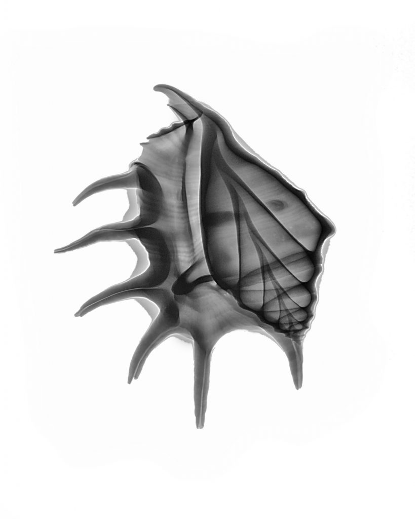
Spider conch (lambis lambis) © Julian Köpke 
Spider conch (lambis lambis) © Julian Köpke -
Orchid X-ray fusion photo

Orchid fusion X-ray photo © Julian Köpke -
Purple Clematis

Three purple Clematis fusion X-ray photo © Julian Köpke -
Purple Clematis

Clematis X-ray photo © Julian Köpke 
Purple Clematis © Julian Köpke 
Purple Clematis X-ray fusion photo © Julian Köpke -
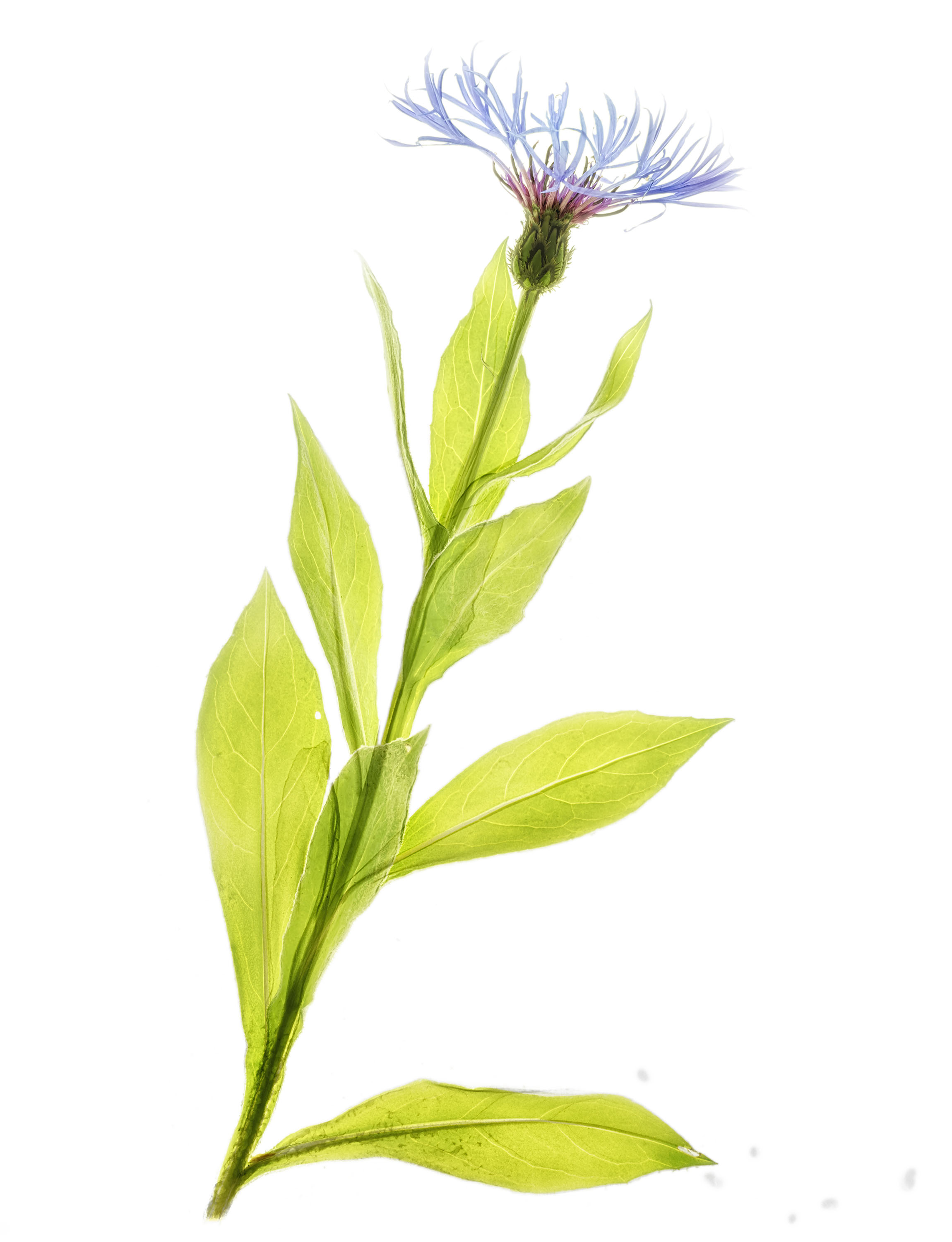
Spring and X-ray fusion photos

Cornflower X-ray fusion photo © Julian Köpke 
Blue aquilegia X-ray fusion photo © Julian Köpke -
Gradients and X-ray tubes

Gradient in an X-ray of 6 red calla lilies © Julian Köpke 
Gradient in an X-ray of 6 red calla lilies, inverted for creative reasons © Julian Köpke


