-
Dahlias fusion X-ray HDR photo

Dahlias using manual HDR in visible light 
Five Dahlias X-ray photo © Julian Köpke 
Dahlias fusion digital X-ray with manual HDR photo in visible light 
Dahlias fusion digital X-ray and manual HDR photo with background © Julian Köpke -
Nautilus shells 3D X-ray photo

Positioning of the three Nautilus shells on the X-ray sensor © Julian Köpke 
Positioning of the three Nautilus shells on the X-ray sensor © Julian Köpke 
Nautilus shell 3D Digital X-ray Photo © Julian Köpke 
Nautilus shell 3D Digital X-ray Photo inverted © Julian Köpke 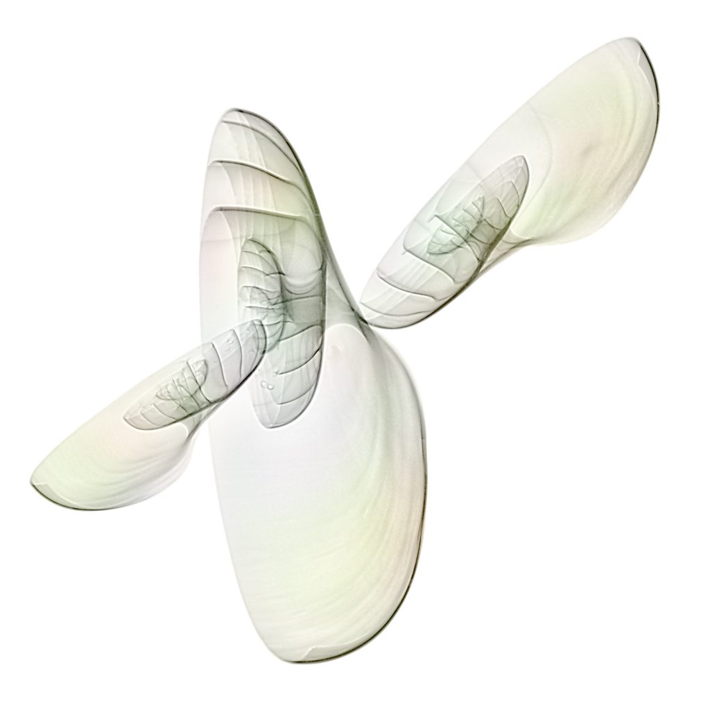
Colorized Nautilus shell 3D Digital X-ray Photo © Julian Köpke 
Nautilus shell 3D Digital X-ray Photo © Julian Köpke 
Nautilus shell 3D Digital X-ray Photo tilted beam © Julian Köpke 
Nautilus shell 3D Digital X-ray Photo tilted beam © Julian Köpke 
Nautilus shell 3D Digital X-ray Photo tilted beam © Julian Köpke -
Sunflower X-ray photos revisited

This sunflower is a fusion photo of X-ray, monochromatic Hα light of the sun converted to BW and a sunflower on a lightbox. © Julian Köpke 
This sunflower is a fusion photo of X-ray, monochromatic Hα light of the sun converted to blue and a sunflower on a lightbox. © Julian Köpke 
Digital X-ray photo of a sunflower (inverted representation). © Julian Köpke -
Shells fusion X-ray photo

Shells fusion X-ray photo © Julian Köpke 
Shells fusion X-ray photo with inversion of the L-channel © Julian Köpke -
Composite of a sunflower: X-ray, light and Hα

This sunflower is a composit of X-ray, monochromatic Hα light of the sun and a sunflower on a lightbox. © Julian Köpke -
Three vetches

Three vetches © Julian Köpke -
X-ray Odyssey

Odyssey © Julian Köpke 
Odyssey (light inversion) © Julian Köpke -
Nautilus and Flowers

Miraculous flowers and nautilus shell © Julian Köpke 
Miraculous flowers © Julian Köpke 
Flowers and Nautilus © Julian Köpke 
Flowers and Nautilus © Julian Köpke 
Nautilus shell as a vase or a vessel („Hansekogge“ or cog) © Julian Köpke 
Positive representation of Nautilus shell as a vase or a vessel („Hansekogge“ or cog) © Julian Köpke 
Coloured Nautilus shell as a vase or a vessel („Hansekogge“ or cog) © Julian Köpke 
Coloured positive representation of a Nautilus shell as a vase or a vessel („Hansekogge“ or cog) © Julian Köpke 
A Nautilus with flowers as Argonauts © Julian Köpke 
A Nautilus with flowers Argonauts © Julian Köpke 
X-Ray positive of a Nautilus with flowers as Argonauts © Julian Köpke 
Colored X-ray positive of a Nautilus with flowers as Argonauts © Julian Köpke -
FAQ: High Dynamic Range


40kV 10mAs 0.031nm 
50kV 2mAs 0.024nm 
60kV 2mAs 0.0207nm 
70kV 10mAs 0.0177nm 
Nautilus X-Ray Energy Compressed © Julian Köpke -
Transparency and Energy in X-Rays

40kV 10mAs 
50kV 2mAs 
60kV 2mAs 
70kV 10mAs 
Nautilus X-Ray Energy HDR © Julian Köpke
Nautilus X-Ray Energy Compressed © Julian Köpke


