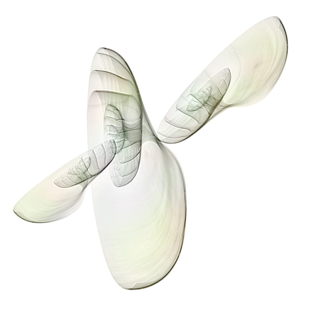
Nautilus shells 3D X-ray photo
How does an X-ray look like with a complete, unsplit specimen of a Nautilus shell ? Will X-rays go through the object ?
My three Nautilus shells I bought in Crete are split specimens. The following approach will give an answer to the question. My composition of my shells is 3-dimensional and in nearly upright position. X-rays were then done with different directions of the radiation to study the effect.


The first image was obtained with radiation coming from the top. The native X-ray representation is with a black background. Historically this was a film negative. Radiologists speak of „transparent“ areas where a film is black. Consequently, white areas are called „opaque“.
The result of radiation coming from the top and slightly tilted shells gives different insights of each shell. The composition looks like a complex mathematical surface or some flying insect.
The inverted (or „positive“) representation is weightless and our mind starts to produce lots of phantasies about the composition.
The effect of colorizing an X-ray is not only graphically. It looks more natural.
The following image was obtained by combining the inverted image with a flat projection of a single shell to a single image. Now one gets an idea of the effect of the beam path.
A tilted beam path shows the a bit more detail of the „wings“. Tilt was about 30 degrees.
Tilt by 45 degrees shows more of a Nautilus as we know it.
Julian Köpke
I like to make things visible the naked eye isn't able to see. That's part of my profession as a radiologist, too.


