-
Tilted Nautilus X-ray photo
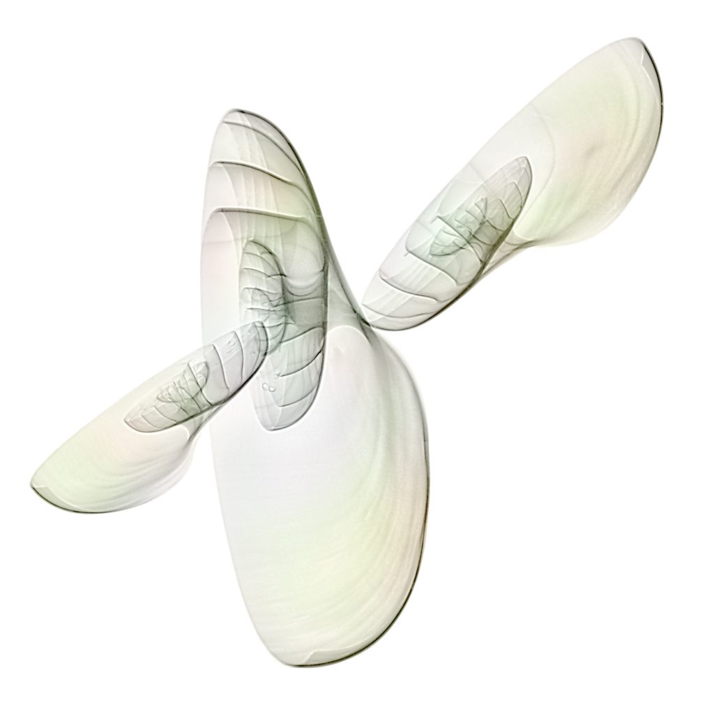
Imagine a Nautilus shell tilted to the surface of the X-ray sensor. The parts close to the sensor are sharp, the distant parts unsharp. Because the X-ray beam creates a central projection. The focal plane is the plane of the sensor, in focus are those parts close to the sensor.
The shell looks like entering the image or leaving it.
-
Nautilus shell X-ray fusion photo of energy levels
Different energies of X-ray radiation mean different transparency of an object. There is an example in my FAQ using a Nautilus shell.
Instead of compressing images of different energies to a single image today I subtracted the 70 kV image of a Nautilus shell from the 40 kV image.
The central parts of the Nautilus shell are more dense and show a significant higher difference. The core of the shell gets shiny. This is how it looks like:
In positive X-ray representation you can compare the results. Left hand is the compressed image of 4 different energy levels, right hand the difference image.
-
Dahlias fusion X-ray HDR photo
Long time ago my friend Harold and I did these X-rays in my practice. There was so much to do. Today was a chance to process the fusion images. Some details can be found in my FAQs.
The manual HDR is already appealing to our eyes.
There is some charm in the X-ray image of the same composition. The hidden parts of the stalks can be clearly seen.
The fusion image of this composition shows both color and hidden structures.
Finished image with a background:
-
Nautilus shells 3D X-ray photo
How does an X-ray look like with a complete, unsplit specimen of a Nautilus shell ? Will X-rays go through the object ?
My three Nautilus shells I bought in Crete are split specimens. The following approach will give an answer to the question. My composition of my shells is 3-dimensional and in nearly upright position. X-rays were then done with different directions of the radiation to study the effect.

Positioning of the three Nautilus shells on the X-ray sensor © Julian Köpke 
Positioning of the three Nautilus shells on the X-ray sensor © Julian Köpke The first image was obtained with radiation coming from the top. The native X-ray representation is with a black background. Historically this was a film negative. Radiologists speak of „transparent“ areas where a film is black. Consequently, white areas are called „opaque“.
The result of radiation coming from the top and slightly tilted shells gives different insights of each shell. The composition looks like a complex mathematical surface or some flying insect.
The inverted (or „positive“) representation is weightless and our mind starts to produce lots of phantasies about the composition.
The effect of colorizing an X-ray is not only graphically. It looks more natural.
The following image was obtained by combining the inverted image with a flat projection of a single shell to a single image. Now one gets an idea of the effect of the beam path.
A tilted beam path shows the a bit more detail of the „wings“. Tilt was about 30 degrees.
Tilt by 45 degrees shows more of a Nautilus as we know it.
-
Sunflower X-ray photos revisited
How to show the sun in the middle of a sunflower ? For astronomers it is quite common to look at the sun in hydrogen alpha light, which is a pure red at 635nm. With artistic eyes, a red center might be overdone.
So I tried two different representations, one in BW that is close to the natural look and feel of a sunflower and one with a light blue in the center as complementary color to the yellow petals.
The surface structure of our sun can be seen like astronomers see it.
There is no photo of the next digital X-ray image of a sunflower with its stalk and a leaf:
-
Shells fusion X-ray photo
Long time I dreamed of this fusion image of shells. Because already on a lightbox some of the shells are transparent and have nice colors. I like the shining through effect very much.
The X-ray image is a compromise of structure and density resolution, depending on the maximum energy the mammography system is able to produce.
Today I’m not at all in a stable state due to a recurrent infection. So I allowed me to do this image instead of hard working.
It is the light inversion in Lab color mode that shows more of a X-ray look and feel. The colors are pretty close to the bright image.
-
Composite of a sunflower: X-ray, light and Hα
In a digital world we can combine different digital sources. This photo of a sunflower is a composit of its X-ray, its photo on a lightbox and monchromatic sunlight at a wavelength of 635nm (Hα light).
In fact: this is an example of an impossible thing. But you may be able to feel the warmth of a sunbeam emerging of the core of the sunflower. And the petals act as prominences.
-
Nautilus and Flowers
How to prepare a X-ray session ? What flowers suit to a Nautilus shell ? Where does color come in ?
I went to my gorgeous florist to have a look what offer she can make during wintertime. My phantasy were spinning around something ethereal or unrealistic. I bought some flowers with respect to their shape.
The Anthuria caught my eye immediately. The Tulip was still closed and got more and more yellow within hours.
All these compositions shown here were made with dual energy X-ray. The lowest energy of the tube is 40kV, which yields with 4 mAs a quite good insight of flowers. For the center of the Nautilus shell, 70kV and 2.5 mAs is more appropriate.
My first composition was a Nautilus taking off a bouquet of flowers. This reminded me of Renaissance engravings full of symbols. I do not feel depressed. The representation as a X-ray positive jsut shows the bouquet.
A more grounded composition is the second with a Nautilus shell moving towards the roots of my bouquet. Hopefully, the plants will survive. The positive representation always needs some extra editing. By just inverting the Blacks and the Whites the Nautilus would be too dark. Our reception cannot be just inverted and feels alright.
With the look-and-feel of old engravings in mind the third composition ist between surreal and a still. It took me some time to mask out the flaws of an original X-ray to get a true black background. Masking can be done iteratively and easily combined with Photoshop. („That’s what Photoshop is made for !“).
Some colorizing was done to overcome missing photographic shots. There was simply no time in my X-ray unit to do both at a time.
My fourth composition is called „The Argonauts“. The Nautilus shell serves as Argo, the legendary fast ship, with its crew, called Argonauts. The colored version is more convenient for our eyes. As before the X-ray positive looks more ethereal.
-
Transparency and Energy in X-Rays
You always need some time to find out the best exposure values for a photo. Same idea holds in X-Ray imaging.
Today I did an x-ray series with my biggest Nautilus shell on a conventional radiography sensor, not a film. Starting from the lowest possible value 40kV an increment of 10 kV up to 70 kV can be seen in the images:
Black regions in the image a transparent, white are opaque. The center of the Nautilus has a loss of structure.
With 50 kV the structure in the center of the Nautilus is better depicted wheras the edge gets more transparent:
Same effect for the center and the edge can be seen with 60 kV:
With 70 kV it’s an exaggeration for the edge and best depiction for the center:
Higher kV means more transparency for denser structures but a loss of structure in transparent areas.
At fixed energy, X-Ray imaging behaves like a shadow related to visible light. When photographing, there is not chance to look through an opaque object. With higher energies, x-rays go through opaque objects and can be collected on a sensor.
Composing the images obtained at different energies is an X-Ray HDR image:
The representation of an X-Ray with white on black is a reminiscence of the film era. Radiologists just looked at the negatives ! Inverting black and white shows the positive image, like a print. Here I show the same image as positive, but rotated and flipped horizontally. Look how ethereal it appears now:
-
X-Ray of Christmas cookies
Thanks to my hard-working father-in-law we enjoy every year phantastic cookies. This year I had to cope with different archiving modalities in mammography due to quality management. This image was an idea to enjoy Christmas in advance with a composition of a Poinsettia. Simple structurizing effects render this image into a sort of cookie smelling painting.


