-
Nautilus shell X-ray fusion photo of energy levels

Energy difference X-ray photo of a Nautilus shell. The image is the difference of a 70kV and a 40 kV image. © Julian Köpke 
Nautilus X-Ray Energy Compressed © Julian Köpke 
Energy difference X-ray photo of a Nautilus shell. The fusion image is the difference of a 70kV and a 40 kV image in X-ray positive representation. © Julian Köpke -
Organ music

Lantern above the fingerboards © Julian Köpke 
Fingerboards of our organ © Julian Köpke 
Organ pipes © Julian Köpke -
Dahlias fusion X-ray HDR photo

Dahlias using manual HDR in visible light 
Five Dahlias X-ray photo © Julian Köpke 
Dahlias fusion digital X-ray with manual HDR photo in visible light 
Dahlias fusion digital X-ray and manual HDR photo with background © Julian Köpke -
Nautilus shells 3D X-ray photo

Positioning of the three Nautilus shells on the X-ray sensor © Julian Köpke 
Positioning of the three Nautilus shells on the X-ray sensor © Julian Köpke 
Nautilus shell 3D Digital X-ray Photo © Julian Köpke 
Nautilus shell 3D Digital X-ray Photo inverted © Julian Köpke 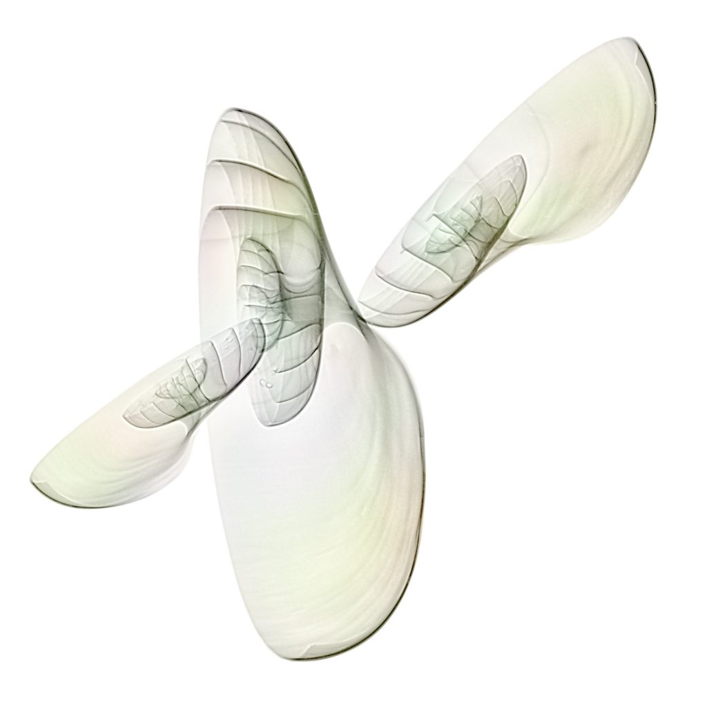
Colorized Nautilus shell 3D Digital X-ray Photo © Julian Köpke 
Nautilus shell 3D Digital X-ray Photo © Julian Köpke 
Nautilus shell 3D Digital X-ray Photo tilted beam © Julian Köpke 
Nautilus shell 3D Digital X-ray Photo tilted beam © Julian Köpke 
Nautilus shell 3D Digital X-ray Photo tilted beam © Julian Köpke -
Sunflower X-ray photos revisited

This sunflower is a fusion photo of X-ray, monochromatic Hα light of the sun converted to BW and a sunflower on a lightbox. © Julian Köpke 
This sunflower is a fusion photo of X-ray, monochromatic Hα light of the sun converted to blue and a sunflower on a lightbox. © Julian Köpke 
Digital X-ray photo of a sunflower (inverted representation). © Julian Köpke -
Shells fusion X-ray photo

Shells fusion X-ray photo © Julian Köpke 
Shells fusion X-ray photo with inversion of the L-channel © Julian Köpke -
Composite of a sunflower: X-ray, light and Hα

This sunflower is a composit of X-ray, monochromatic Hα light of the sun and a sunflower on a lightbox. © Julian Köpke -
Primroses

Purple Primroses © Julian Köpke 
Primroses II © Julian Köpke 
Primroses I © Julian Köpke -
Three vetches

Three vetches © Julian Köpke -
End of wintertime

Physalis after winter has gone. © Julian Köpke


