-
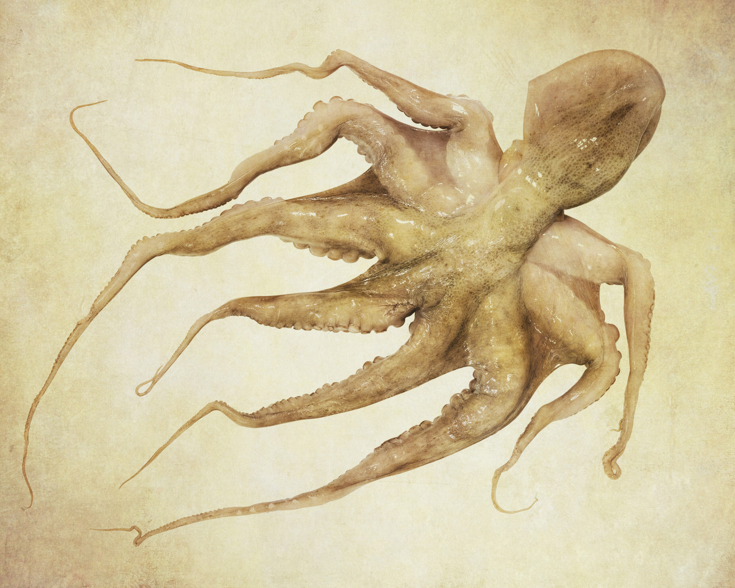
Pulpo
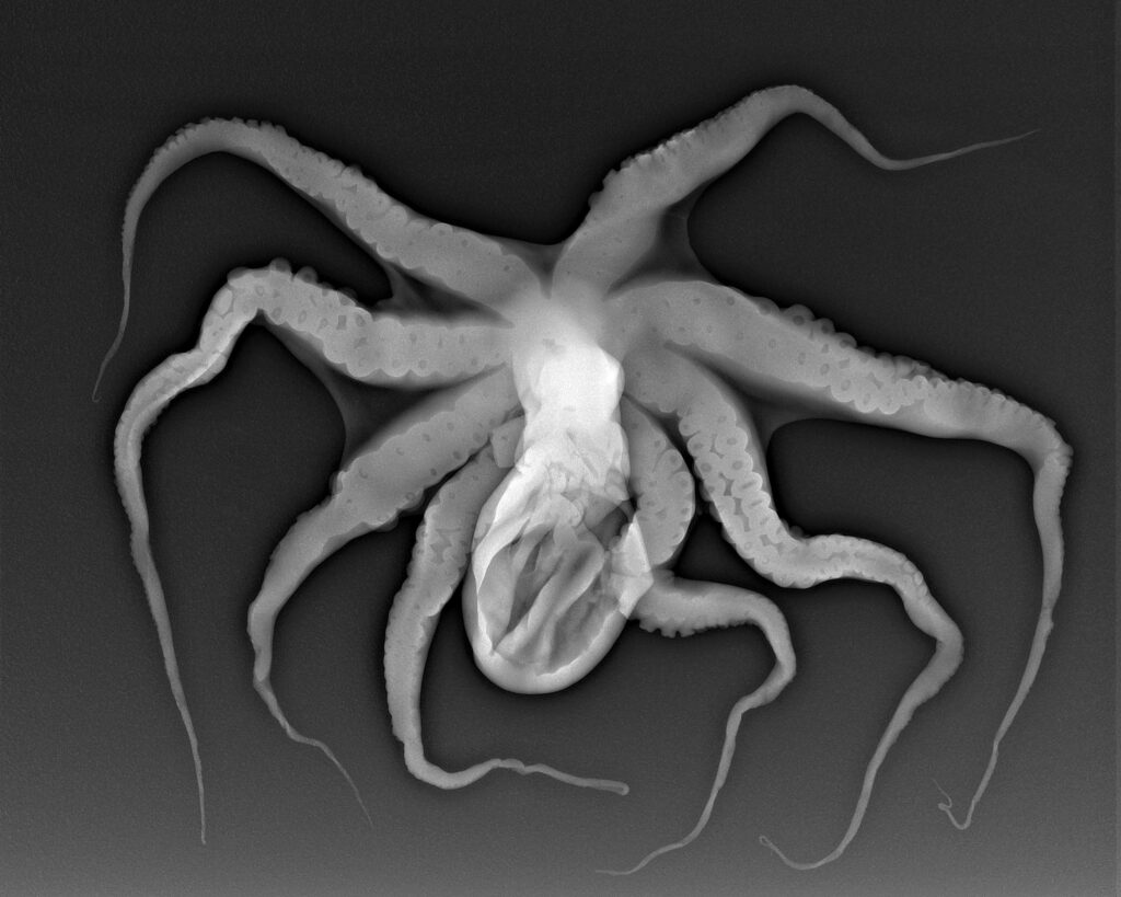
Pulpo © Julian Köpke 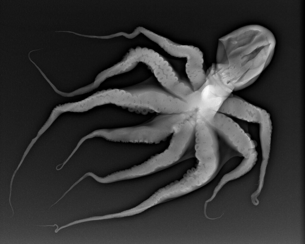
Pulpo © Julian Köpke 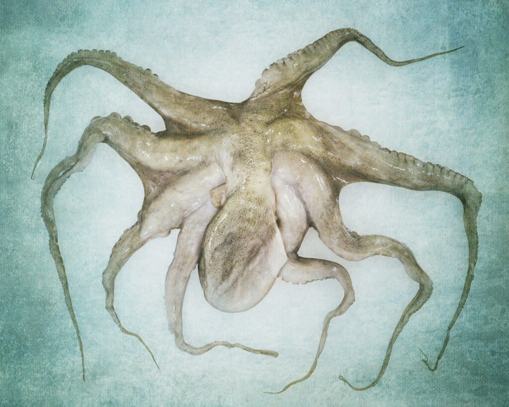
Pulpo © Julian Köpke 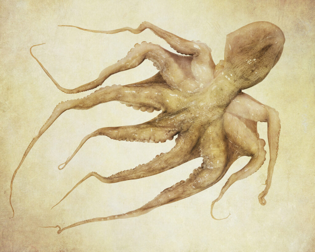
Pulpo © Julian Köpke -
X-ray of vegetables and boxes
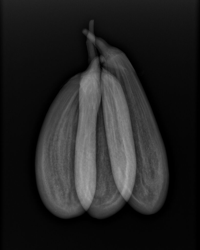
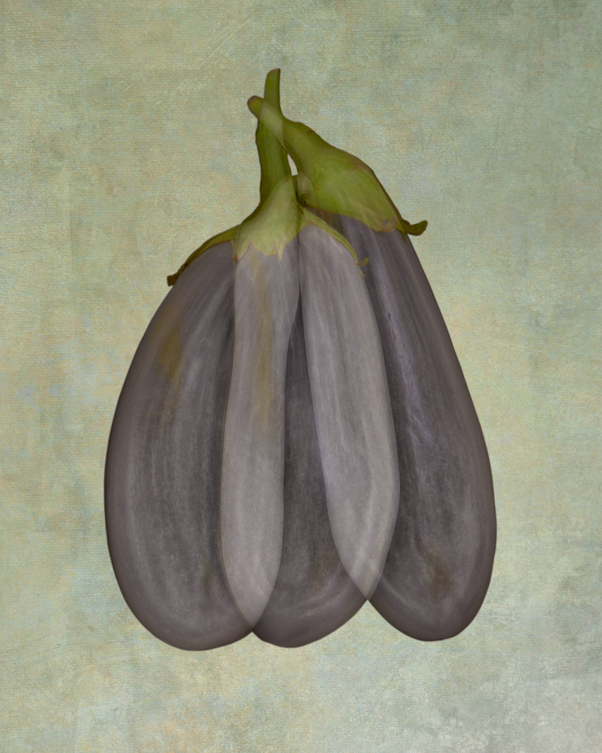
X-ray of aubergines © Julian Köpke 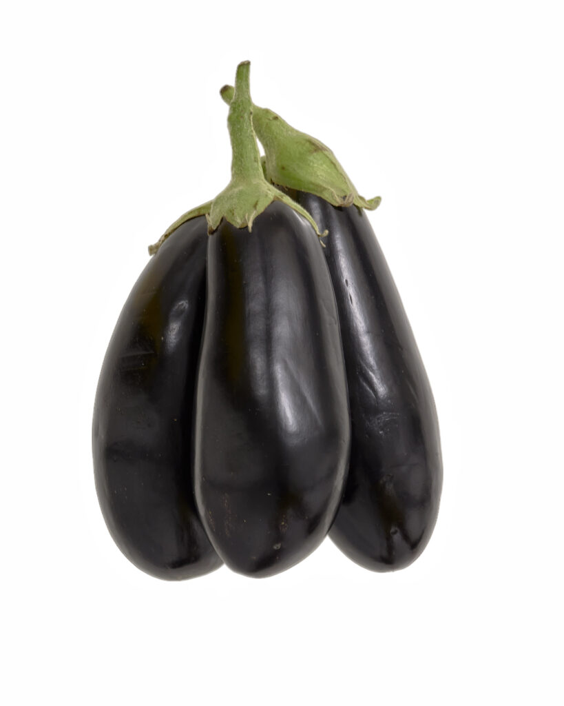
Aubergines © Julian Köpke 
Aubergines X-ray fusion photography © Julian Köpke 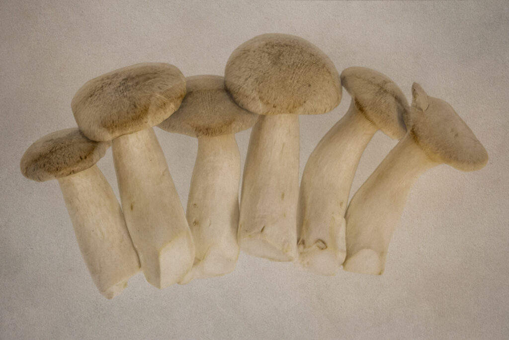
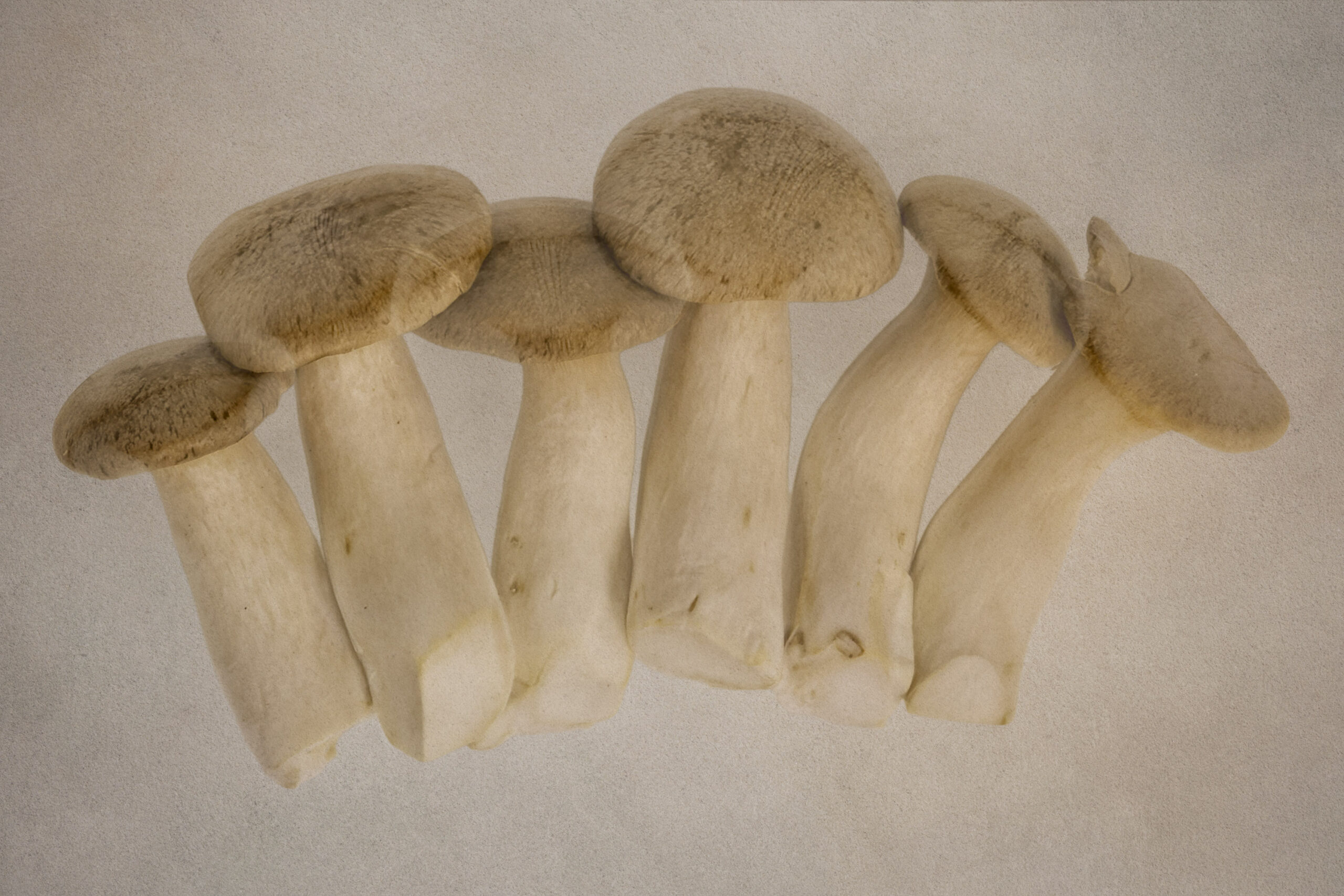
Mushrooms X-ray fusion photography © Julian Köpke 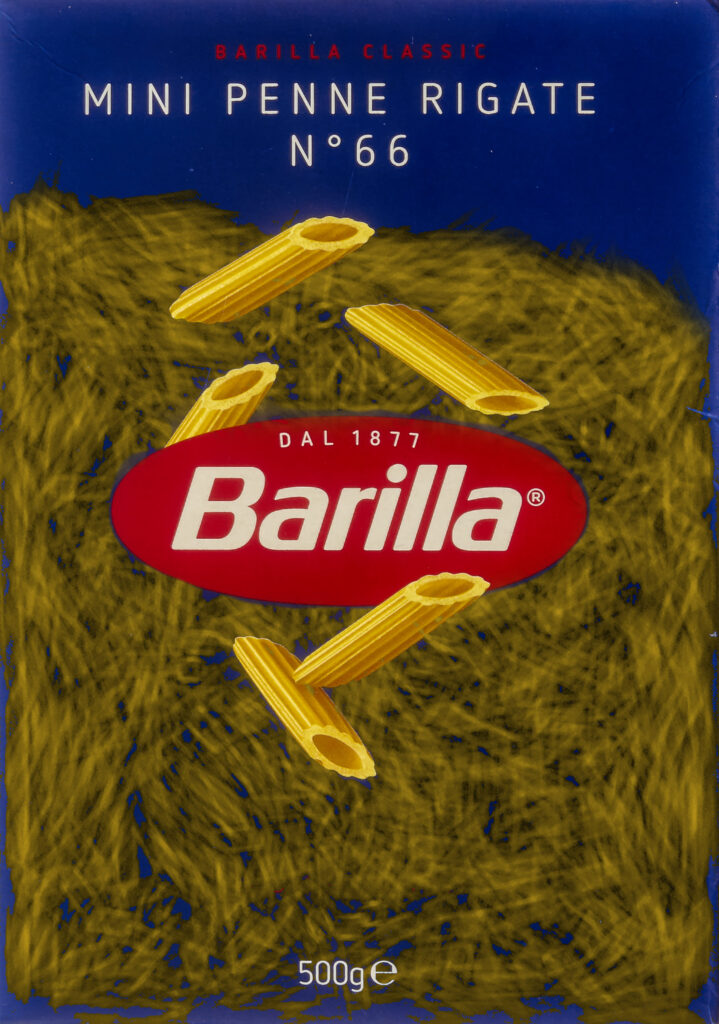
Mini Penne Rigate n. 66 © Julian Köpke 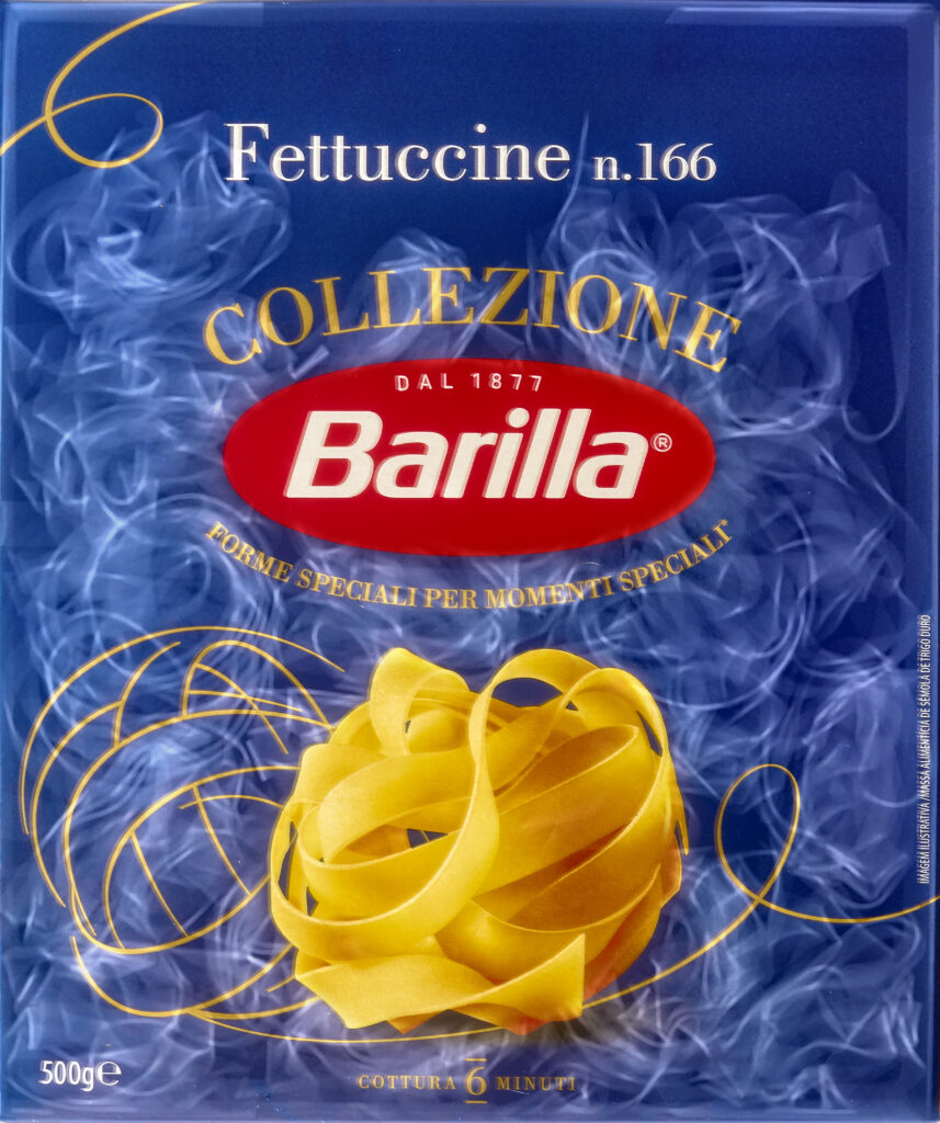
Fettuccine n.166 X-ray fusion photography © Julian Köpke -
Colorizing an X-ray

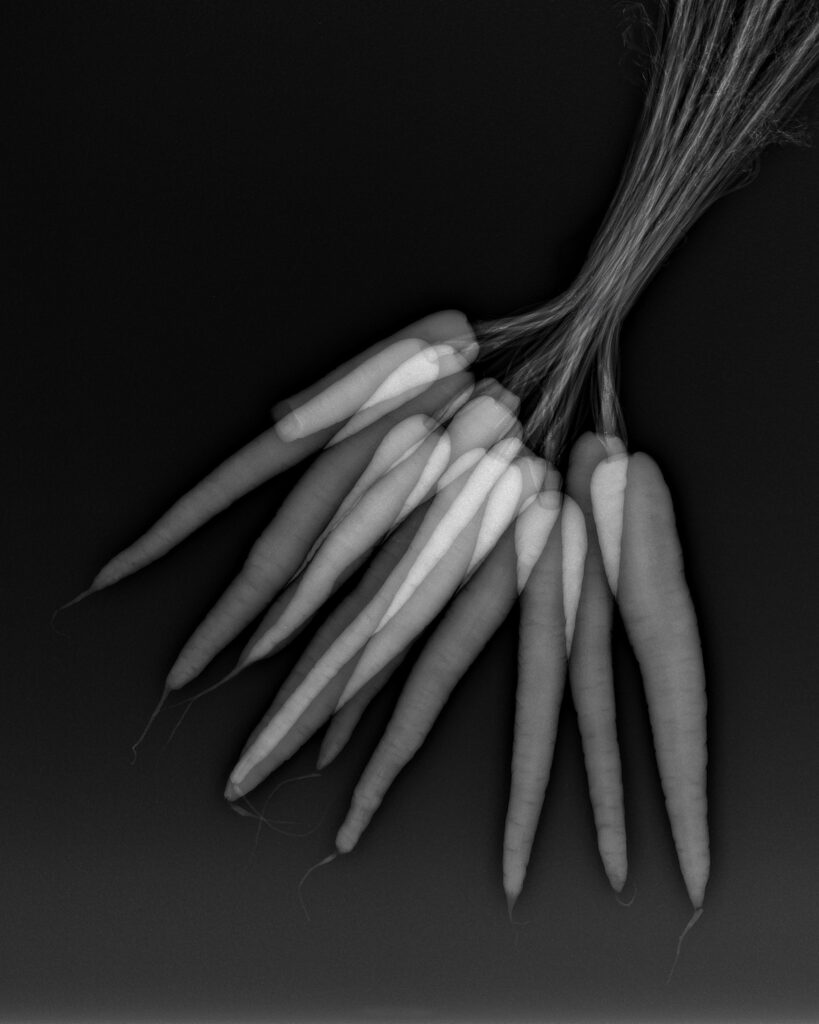
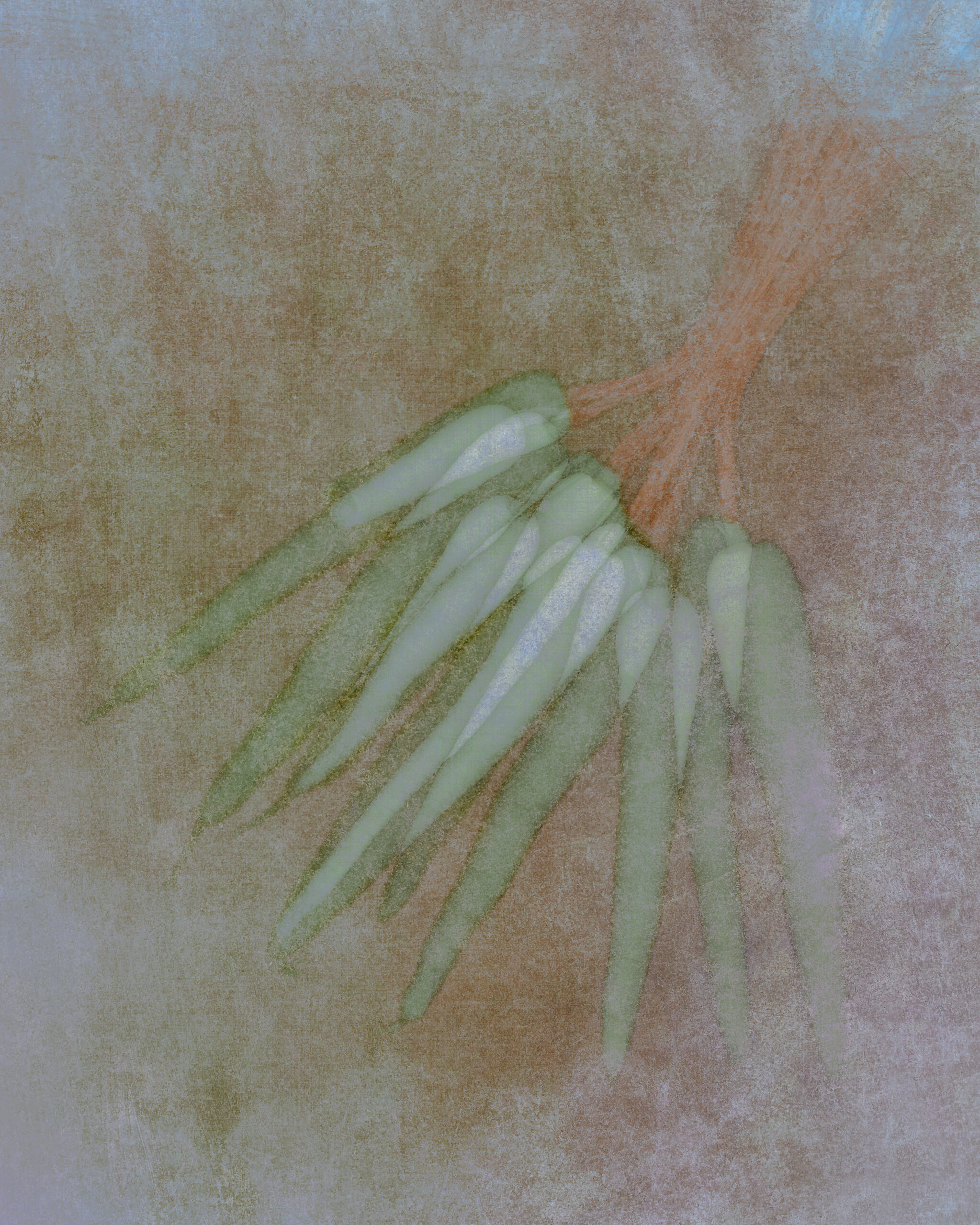
Bunch of carrots © Julian Köpke 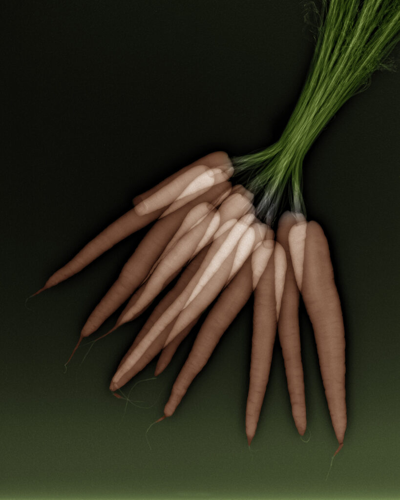
Bunch of carrots texturized © Julian Köpke 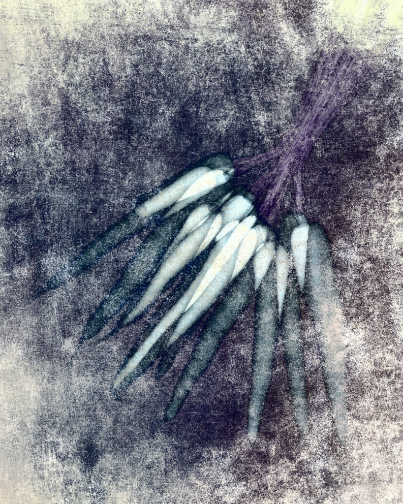
Bunch of carrots © Julian Köpke 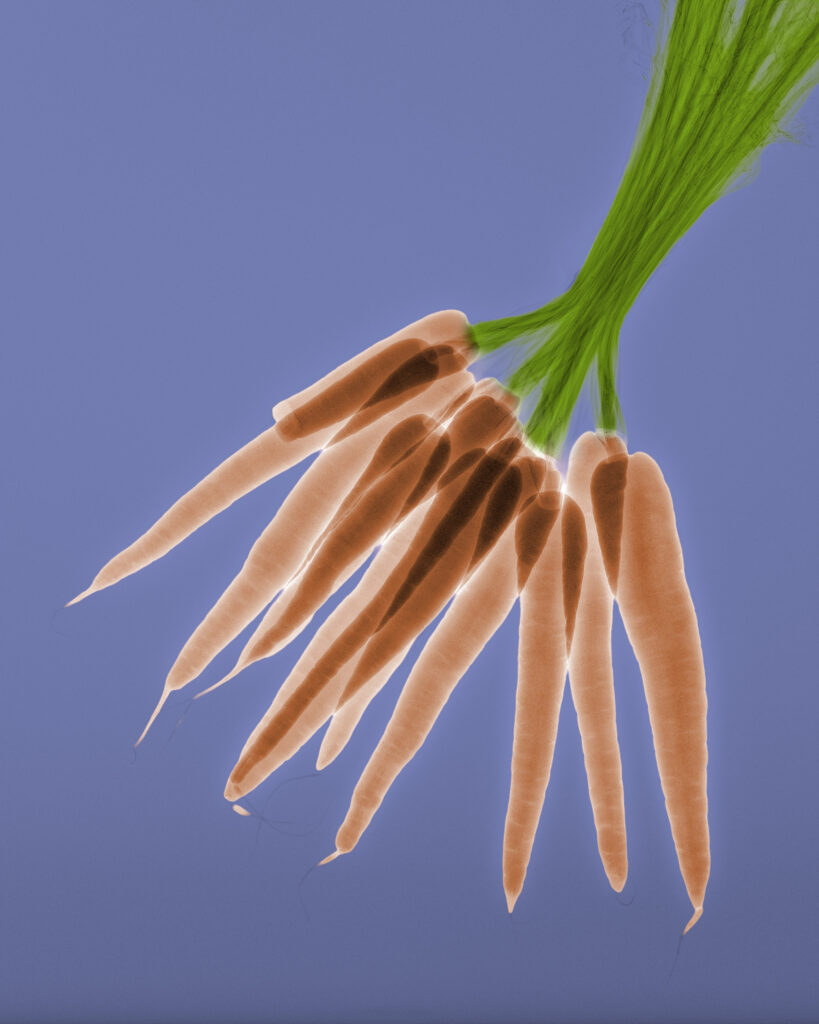
Bunch of carrots © Julian Köpke 
Bunch of carrots © Julian Köpke 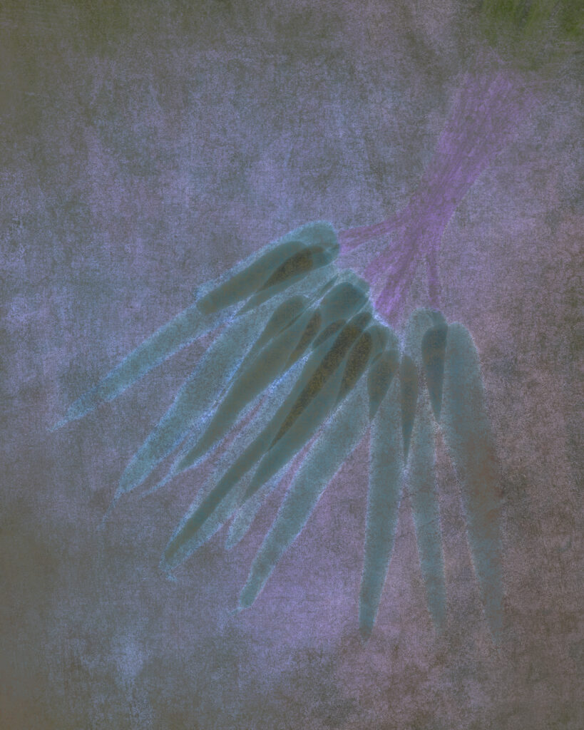
Bunch of carrots © Julian Köpke -
Varieties of fusion images using X-ray

Pink lily X-ray fusion photography © Julian Köpke 
White Campanula X-ray fusion photography © Julian Köpke 
Amaryllis X-ray mammography fusion photography texturized © Julian Köpke 
Fusion X-ray photo Calla lilies IV. Black background using Lab inversion. © Julian Köpke 
X-ray fusion photo snail shells composition III © Julian Köpke 
X-ray fusion photo snail shells composition III © Julian Köpke 
Sea bream (Dorade) - X-ray fusion photography © Julian Köpke 
Trout (Forelle) - X-ray fusion photography © Julian Köpke -
Transparencies

Two lily blossoms © Julian Köpke -
Structures
-
Fusion Imaging of Flowers
-
X-ray of Flowers
-
Flowers
-
FAQ: X-rays


