-
Spider conch X-ray fusion photo

Fusion image spider conch X-ray and photo seen from the bottom © Julian Köpke 
Fusion image spider conch (lambis lambis) X-ray and photo seen from above © Julian Köpke 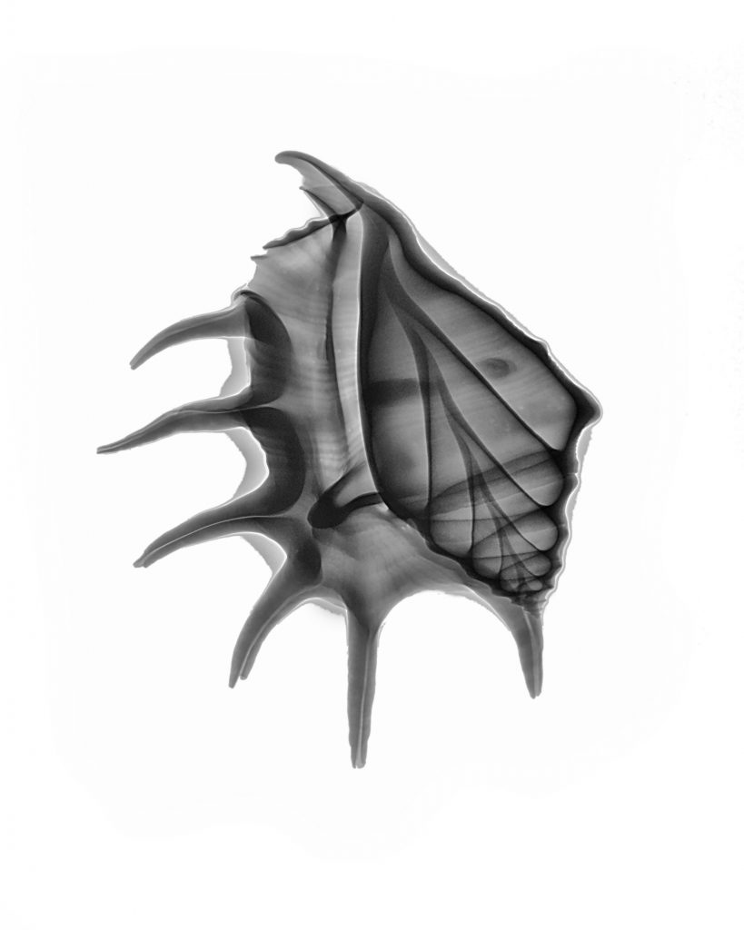
Spider conch (lambis lambis) © Julian Köpke 
Spider conch (lambis lambis) © Julian Köpke -
X-ray Odyssey

Odyssey © Julian Köpke 
Odyssey (light inversion) © Julian Köpke -
Nautilus and Flowers

Miraculous flowers and nautilus shell © Julian Köpke 
Miraculous flowers © Julian Köpke 
Flowers and Nautilus © Julian Köpke 
Flowers and Nautilus © Julian Köpke 
Nautilus shell as a vase or a vessel („Hansekogge“ or cog) © Julian Köpke 
Positive representation of Nautilus shell as a vase or a vessel („Hansekogge“ or cog) © Julian Köpke 
Coloured Nautilus shell as a vase or a vessel („Hansekogge“ or cog) © Julian Köpke 
Coloured positive representation of a Nautilus shell as a vase or a vessel („Hansekogge“ or cog) © Julian Köpke 
A Nautilus with flowers as Argonauts © Julian Köpke 
A Nautilus with flowers Argonauts © Julian Köpke 
X-Ray positive of a Nautilus with flowers as Argonauts © Julian Köpke 
Colored X-ray positive of a Nautilus with flowers as Argonauts © Julian Köpke -
FAQ: High Dynamic Range


40kV 10mAs 0.031nm 
50kV 2mAs 0.024nm 
60kV 2mAs 0.0207nm 
70kV 10mAs 0.0177nm 
Nautilus X-Ray Energy Compressed © Julian Köpke -
Mediterranean creatures on a lightbox

Cretean Snail © Julian Köpke 
Mediterranean Snail II © Julian Köpke 
Three Nautilus shells with light inverison © Julian Köpke


