-
Heucheria leaf on a light box

Heucheria leaf © Julian Köpke 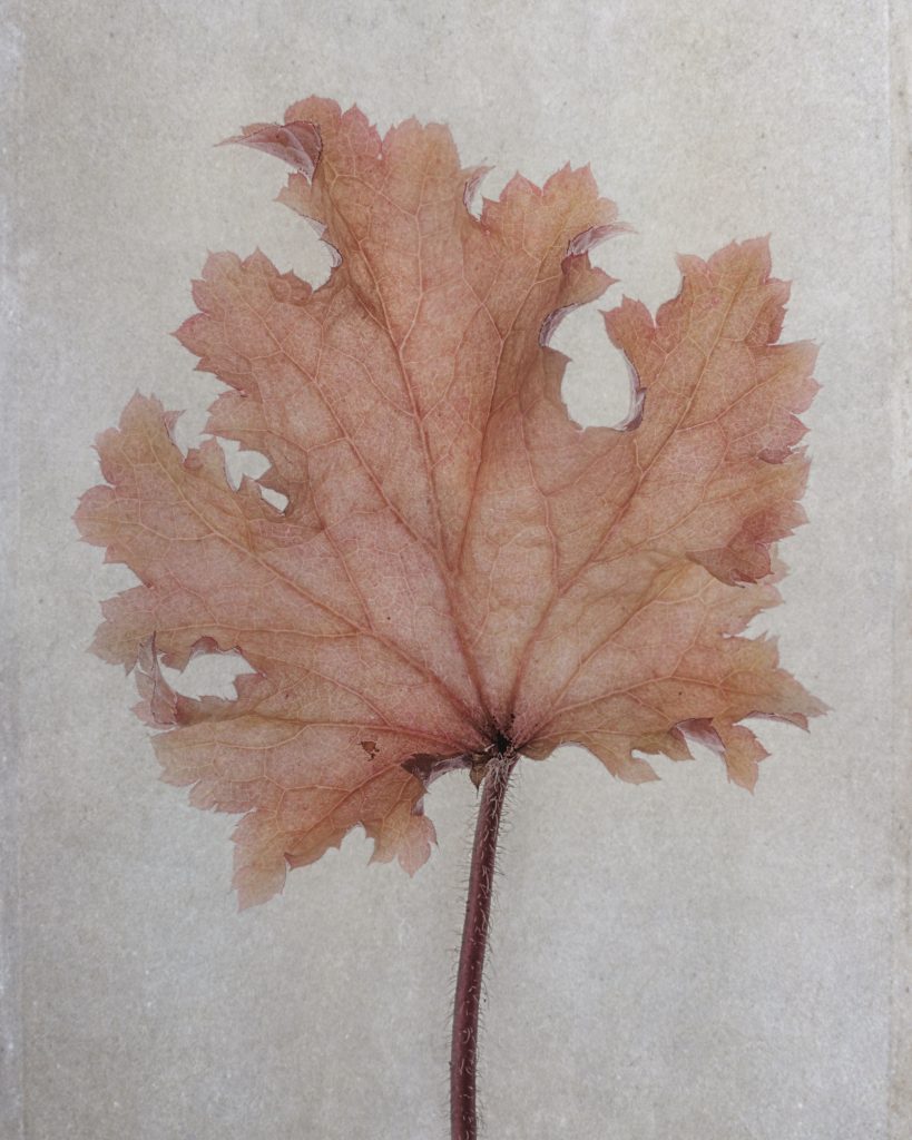
Heucheria leaf © Julian Köpke 
Heucheria leaf © Julian Köpke -
Orchid X-ray fusion photo

Orchid fusion X-ray photo © Julian Köpke -
Purple Clematis

Three purple Clematis fusion X-ray photo © Julian Köpke -
Purple Clematis

Clematis X-ray photo © Julian Köpke 
Purple Clematis © Julian Köpke 
Purple Clematis X-ray fusion photo © Julian Köpke -
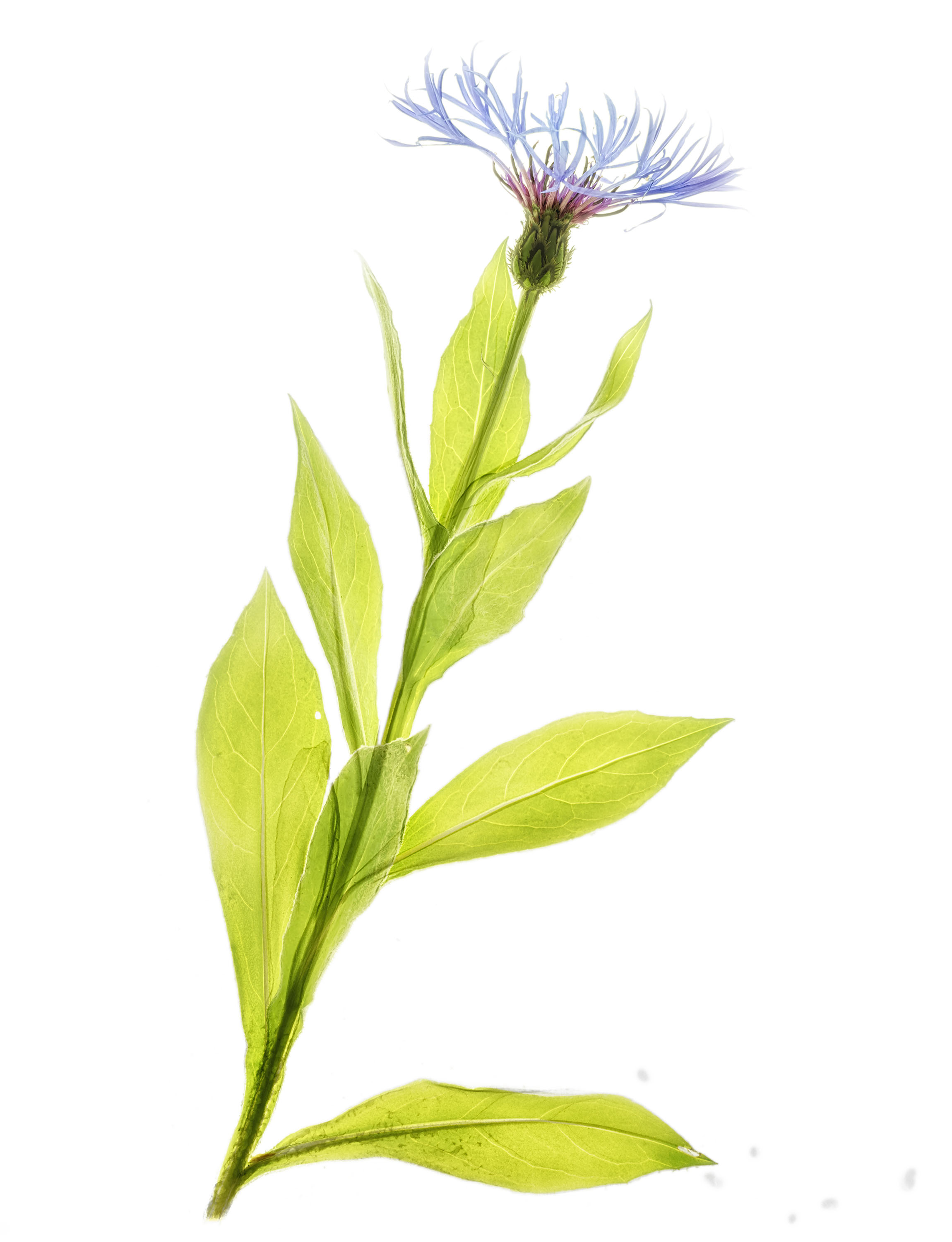
Spring and X-ray fusion photos

Cornflower X-ray fusion photo © Julian Köpke 
Blue aquilegia X-ray fusion photo © Julian Köpke -
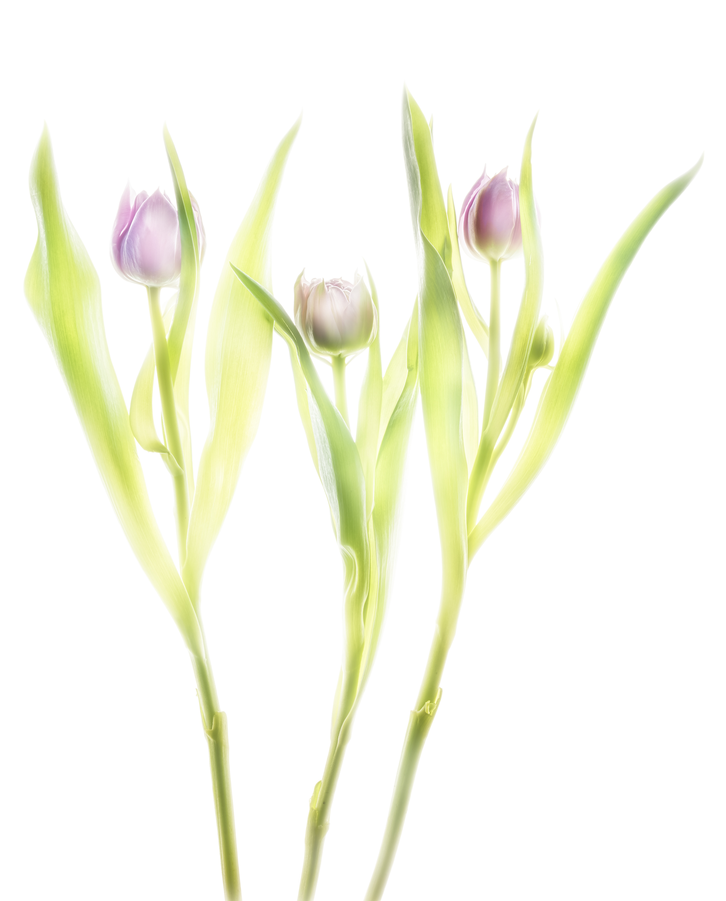
Fusion X-ray of tulips

X-ray three tulips © Julian Köpke 
Three purple tulips HDR photo © Julian Köpke 
Three purple tulips fusion X-ray photo © Julian Köpke -
Radiating Beauty: Creating a new photographic form with fusion X-Ray images

X-ray fusion image of a Gloriosa lilly © Julian Köpke 
This sunflower is a composit of X-ray, monochromatic Hα light of the sun and a sunflower on a lightbox. © Julian Köpke -
X-ray fusion photo of a Nautilus

Nautilus shell manual HDR photo on a light box © Julian Köpke 
Nautilus X-Ray Energy Compressed © Julian Köpke 
Nautilus shell fusion X-ray photo and manual HDR photo on a light box. © Julian Köpke -
Dahlias fusion X-ray HDR photo

Dahlias using manual HDR in visible light 
Five Dahlias X-ray photo © Julian Köpke 
Dahlias fusion digital X-ray with manual HDR photo in visible light 
Dahlias fusion digital X-ray and manual HDR photo with background © Julian Köpke -
Shells fusion X-ray photo

Shells fusion X-ray photo © Julian Köpke 
Shells fusion X-ray photo with inversion of the L-channel © Julian Köpke