-
Purple Clematis
Long lasting blossoms, turning up every year: my purple clematis in our garden.
It was my third X-ray session with flowers this week. Third fusion imaging attempt. After blue cornflower and blue aquilegia now a purple clematis. Big data on my hard disk.
Today we did it with mammography at 30 kV and 50 mAs. Lower noise ! Here is the positive representation of a single clematis:
I processed the lightbox HighKey series with a mask. There was a shift of 2 or 3 pixels from the lightest to the darker images. So I processed everything a second time to compensate for the shift. The HDR image shows a cut stalk. Photoshop is made for this.
The stalk can be lengthened like in the preceding X-ray. The fusion image shows hidden leaves, the core of the blossom and stalks much better:
The flower looks pretty fragile now, close to its natural appearance.
-
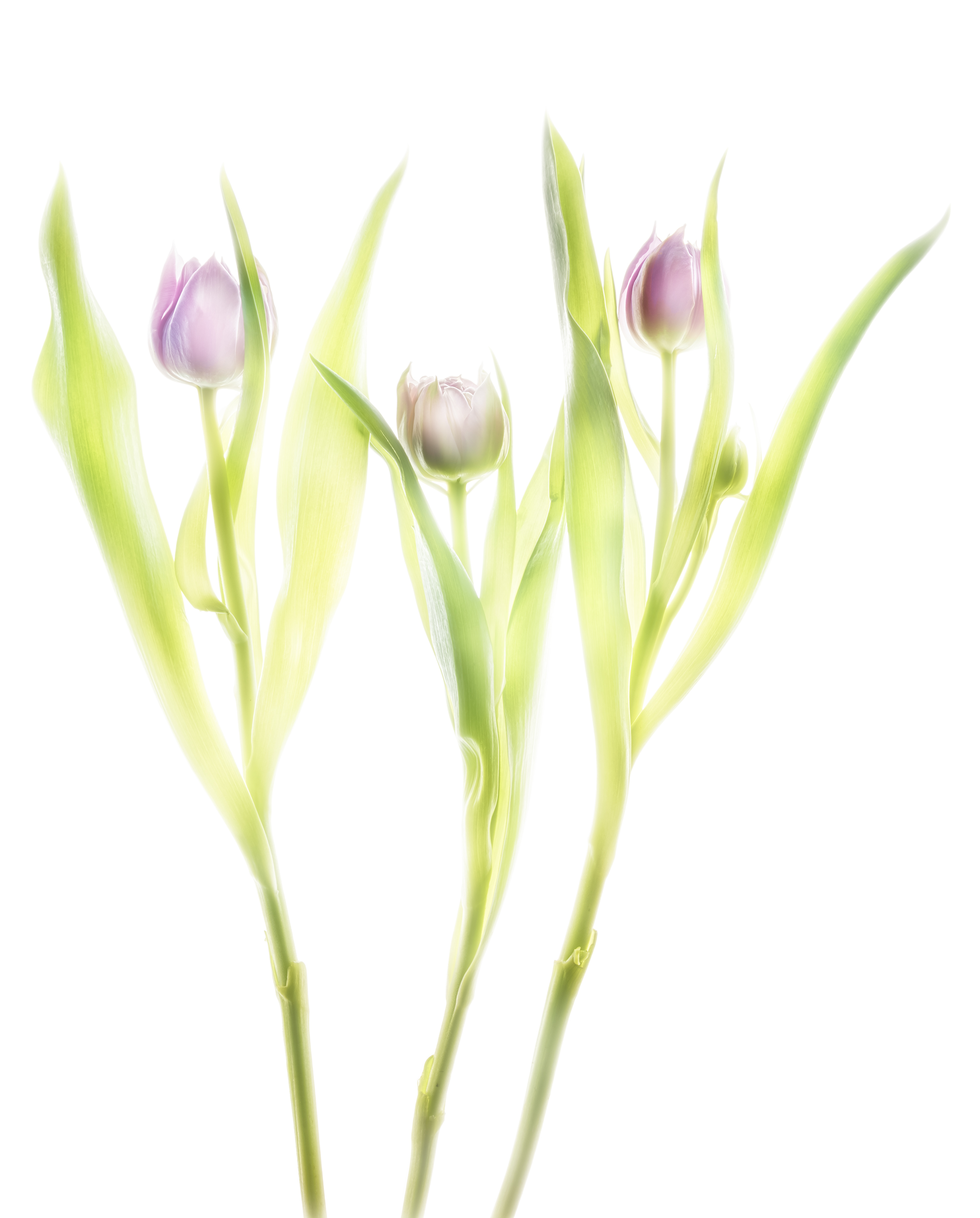
Fusion X-ray of tulips
Fusion imaging is beauty made of composite X-ray images and HDR images on a light box. The primary question is what energy fits best for flowers. To my experience 40 kV is often suitable. But: the proof of the pudding is in the eating.
Mammography systems start e.g. from 20 kV and reach 39 kV. The sensor is up to 24cm x 30cm. Conventional systems start from 40 kV and reach 125 kV. The sensor is up to 43cm x 43cm what makes them more attractive to floral compositions.
The higher resolution and the lower energies of a mammography will suit better for transparent objects. But the spatial limit of a composition (which is 24cm x 30cm) might put hard restrictions on the artist.
Floral compositions have more creative space with a bigger sensor. But the X-ray tube starts with 40 kV and this might lead to overexposure of tender structures.
Thus I performed today more than ten compositions to study this relation.
After four exposure of three tulips I found this composition with four dense blossoms attractive to go further. The composition might somehow resemble to a sketch of three angels. The image is nice due to very soft edges of their „wings“, technically blown out portions in the image. The inner structure of the nearly closed blossoms is well resolved. The stalks serve as „body“. There is no advantage with higher energies.
The same composition was done immediately after the X-ray as a bracketing series on a lightbox. After returning I processed a manual HDR, the colors not to warm.
The final fusion image is a composite of the preceding two images. Compared to the lightbox photo, the hidden stalks reappear naturally, the inner petals are outlined like a sketch.
-
Radiating Beauty: Creating a new photographic form with fusion X-Ray images
The shapes and forms are recognizable, yet the level of detail is deeper than the human eye can normally perceive: Leaves appear minutely laced and surfaces are impossibly intricate, somewhere between translucent and opaque. Welcome to the captivating work of photographer Harold Davis and radiologist Dr. Julian Köpke, who combine their skill, passion, and vision to create stunning X-ray photography and pioneering fusion images. Read more on the Pixsy blog (article by Natalie Holmes).
This nice article was posted today to share the fascination of our common work on fusion X-ray images using a light box manual HDR photo of flowers and their X-ray.
Our X-ray data are the same, our photographic data a nearly the same: Harold used a Nikon D850 and I used a Nikon D810A, which is modified for astrophotography. Our common lens was a Zeiss Makro-Planar T* 2/50 ZF.2.
We have some techniques and some principles in common, yet we are different individuals with different results. The next image is partly inspired by Harold’s version. Blue is the complementary color to yellow and fits nicely into the petals. The red color in the center is an image of the sun in monochromatic Hα light using a Fabry-Perot-Interferometer. So this image is a triple fusion image of three different light sources ! If you look closer at 2pm in the center, there are two sunspots.
-
Shells fusion X-ray photo
Long time I dreamed of this fusion image of shells. Because already on a lightbox some of the shells are transparent and have nice colors. I like the shining through effect very much.
The X-ray image is a compromise of structure and density resolution, depending on the maximum energy the mammography system is able to produce.
Today I’m not at all in a stable state due to a recurrent infection. So I allowed me to do this image instead of hard working.
It is the light inversion in Lab color mode that shows more of a X-ray look and feel. The colors are pretty close to the bright image.
-
Composite of a sunflower: X-ray, light and Hα
In a digital world we can combine different digital sources. This photo of a sunflower is a composit of its X-ray, its photo on a lightbox and monchromatic sunlight at a wavelength of 635nm (Hα light).
In fact: this is an example of an impossible thing. But you may be able to feel the warmth of a sunbeam emerging of the core of the sunflower. And the petals act as prominences.
-
Primroses
I felt very much attracted by these primroses. They were close to purple and red and I could see them already as a beautiful print.
But how photographing them on a lightbox ? They always toppled over. Many efforts were useless. Blossoms tend to move, always.
On this photograph I put the blossoms top-down. Because any arrangement could be done then. It works !
A different color show the orange primroses. Composition with or without leaves ? Without gives more the impression of a painting.
-
Three vetches
X-ray images give an insight into the inner (or hidden) structure of a flower. HDR images on a light box are quite close to this.
Today I wanted to show the softness of petals and went to my dealer. She sold me three vetches, not really expensive for the purpose.
This is my third composition today of the three vetches on my lightbox. The play of the light in the petals resembles to some extent X-ray images.
-
End of wintertime
Our weather is more and more weird. Today was the second day with a warm sun and a blue sky. Nights are getting pretty cold, days up to 25 degrees Celsius.
Cleaning up our garden led us to some old physalis which were a little more than a skeleton. In autumn these fruits look like lanterns, now they resemble an X-ray.
I did this shot on a lightbox using manual HDR technique.In Lab color mode I obtained this image with a pur black background.
It’s an exoskeleton for the fruit inside which remains that way without bruises.
-
Mediterranean creatures on a lightbox
Today I put some tests on my cretean purchases from last September to evaluate their potential of being subject to fusion imaging. I bought three Nautilus shells and two sea snails, holding them in the store against the sun to check their transparency. My untidy studio accommodated these precious stones under quite a bunch of something.
The best representation is with a black background, i.e. with inverted L-channel in Lab colors. With a black background a soft shining light appears in the objects.
This snail has a shape a triangle and resembles a bear claw or an Apollo capsule in the late Sixties. The translucency is very little.
The following snail has a classic shape. With the black background it resembles a galaxy in outer space.
My first attempt with the Nautilus shells led me to a copper-like color representation with a single shot image. Lab colors is the key to this color and light distribution. Very attractive is the fact of two shells turning right and one left. Why did I wait so long to make this image ? Why do we miss important opportunities ?
-
X-ray fusion images
3. November 2018 /