-
Snail shells X-ray fusion photos
A friend handed me out some snail shells that he had in mind for a long time to lend me. Eventually, he found 5 beautiful shells when cleaning up the basement.
The effect of the images depends strongly on the post-processing. Some of the results may not be combined in one presentation.
Here I show three images of them as dark jewels with an intrinsic undefinable light. Maybe, we are thousand miles below sea level.
Fusion imaging works with a light box. Without, too. It depends on your subject. The light images were taken with a Leica Q, pointing just in the same direction as the X-rays from below of the X-ray tube. The resolution and technology is completely sufficient for the color use.
I designed a new composition, which should allow me to have different positions of the shells in space. The surrounding snail shells serve as supports.
I wanted to take the yellow, quit radiopaque snail shell from above. So I had to rearrange the snail shells once more.
When looking at my flickr stream you may find other representations in the preceding neighborhood of this image.
-
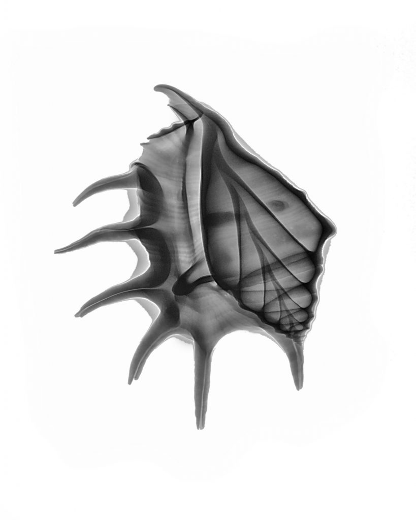
Spider conch X-ray fusion photo
A friend gave me a shell of a spider conch to make more fusion images. The scientific name of the spider conch is lambis lambis and it is a sea snail. There is a nice Wikipedia article on it.
The hard shell with a lot of radiopaque lime made me doubt the success of my X-rays. On top, my first attempt at a HighKey image wasn’t really convincing. Only the combination of a normal photography for the color, a HighKey image for a transparency effect together with the X-ray image resulted in nice image.
The X-ray image appear less lively, but full of formal power. The orientation of the animal is conveyed by the photographically reproduced color. There are only minimal hints wich orientation the X-ray has.
These are the corresponding X-ray images:
-
Mediterranean creatures on a lightbox
Today I put some tests on my cretean purchases from last September to evaluate their potential of being subject to fusion imaging. I bought three Nautilus shells and two sea snails, holding them in the store against the sun to check their transparency. My untidy studio accommodated these precious stones under quite a bunch of something.
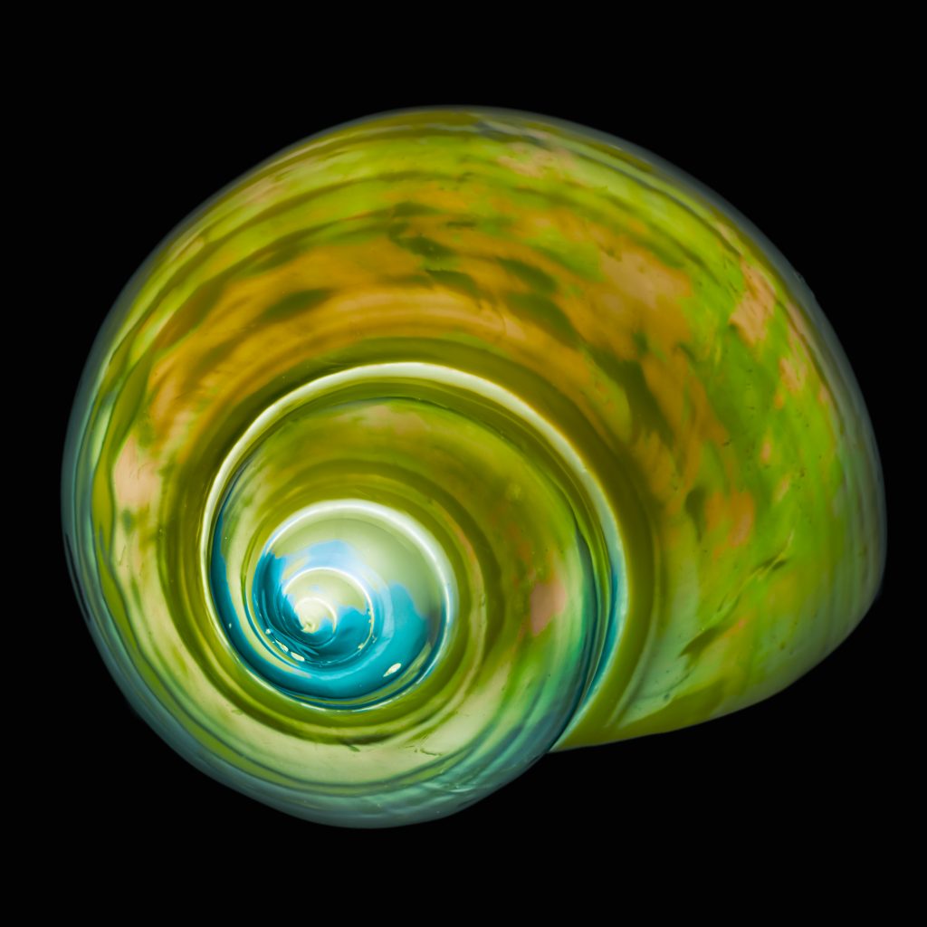
The best representation is with a black background, i.e. with inverted L-channel in Lab colors. With a black background a soft shining light appears in the objects.
This snail has a shape a triangle and resembles a bear claw or an Apollo capsule in the late Sixties. The translucency is very little.
The following snail has a classic shape. With the black background it resembles a galaxy in outer space.
My first attempt with the Nautilus shells led me to a copper-like color representation with a single shot image. Lab colors is the key to this color and light distribution. Very attractive is the fact of two shells turning right and one left. Why did I wait so long to make this image ? Why do we miss important opportunities ?


