-
Fusion X-ray of tulips
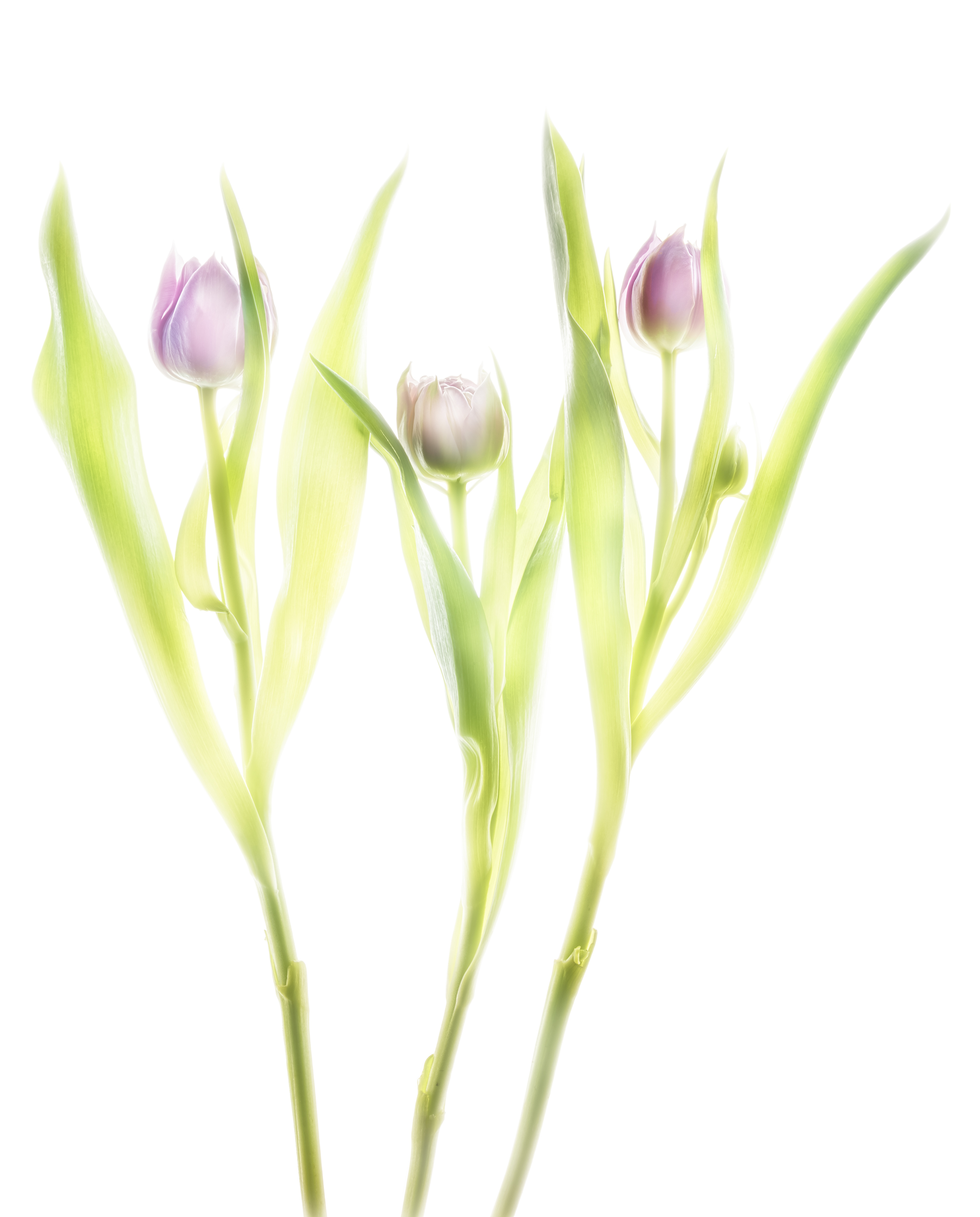
Fusion imaging is beauty made of composite X-ray images and HDR images on a light box. The primary question is what energy fits best for flowers. To my experience 40 kV is often suitable. But: the proof of the pudding is in the eating.
Mammography systems start e.g. from 20 kV and reach 39 kV. The sensor is up to 24cm x 30cm. Conventional systems start from 40 kV and reach 125 kV. The sensor is up to 43cm x 43cm what makes them more attractive to floral compositions.
The higher resolution and the lower energies of a mammography will suit better for transparent objects. But the spatial limit of a composition (which is 24cm x 30cm) might put hard restrictions on the artist.
Floral compositions have more creative space with a bigger sensor. But the X-ray tube starts with 40 kV and this might lead to overexposure of tender structures.
Thus I performed today more than ten compositions to study this relation.
After four exposure of three tulips I found this composition with four dense blossoms attractive to go further. The composition might somehow resemble to a sketch of three angels. The image is nice due to very soft edges of their „wings“, technically blown out portions in the image. The inner structure of the nearly closed blossoms is well resolved. The stalks serve as „body“. There is no advantage with higher energies.
The same composition was done immediately after the X-ray as a bracketing series on a lightbox. After returning I processed a manual HDR, the colors not to warm.
The final fusion image is a composite of the preceding two images. Compared to the lightbox photo, the hidden stalks reappear naturally, the inner petals are outlined like a sketch.
-
Radiating Beauty: Creating a new photographic form with fusion X-Ray images
The shapes and forms are recognizable, yet the level of detail is deeper than the human eye can normally perceive: Leaves appear minutely laced and surfaces are impossibly intricate, somewhere between translucent and opaque. Welcome to the captivating work of photographer Harold Davis and radiologist Dr. Julian Köpke, who combine their skill, passion, and vision to create stunning X-ray photography and pioneering fusion images. Read more on the Pixsy blog (article by Natalie Holmes).
This nice article was posted today to share the fascination of our common work on fusion X-ray images using a light box manual HDR photo of flowers and their X-ray.
Our X-ray data are the same, our photographic data a nearly the same: Harold used a Nikon D850 and I used a Nikon D810A, which is modified for astrophotography. Our common lens was a Zeiss Makro-Planar T* 2/50 ZF.2.
We have some techniques and some principles in common, yet we are different individuals with different results. The next image is partly inspired by Harold’s version. Blue is the complementary color to yellow and fits nicely into the petals. The red color in the center is an image of the sun in monochromatic Hα light using a Fabry-Perot-Interferometer. So this image is a triple fusion image of three different light sources ! If you look closer at 2pm in the center, there are two sunspots.
-
X-ray fusion photo of a Nautilus
Fusion imaging can be done retrospective. My split Nautilus shell on a light box rendered with manual HDR shows already a nice structure of the inner parts.
The X-ray obtained a couple of days earlier easily fits onto the HDR with not a big deal of processing.
The meaning of the fusion image may be different to the flowers. But it’s feasible to do it retrospectively.
-
Nautilus shell X-ray fusion photo of energy levels
Different energies of X-ray radiation mean different transparency of an object. There is an example in my FAQ using a Nautilus shell.
Instead of compressing images of different energies to a single image today I subtracted the 70 kV image of a Nautilus shell from the 40 kV image.
The central parts of the Nautilus shell are more dense and show a significant higher difference. The core of the shell gets shiny. This is how it looks like:
In positive X-ray representation you can compare the results. Left hand is the compressed image of 4 different energy levels, right hand the difference image.
-
Dahlias fusion X-ray HDR photo
Long time ago my friend Harold and I did these X-rays in my practice. There was so much to do. Today was a chance to process the fusion images. Some details can be found in my FAQs.
The manual HDR is already appealing to our eyes.
There is some charm in the X-ray image of the same composition. The hidden parts of the stalks can be clearly seen.
The fusion image of this composition shows both color and hidden structures.
Finished image with a background:
-
Blog
3. November 2018 / -
FAQ: Fusion imaging
3. November 2018 /Explanation of the idea
Fusion imaging is a child of the digital era of mapping structures. Before image fusion was used in diagnostic radiology, astronomers used it to extract new insights from our universe. Fusion imaging of flowers can be beautiful. And, maybe, it’s a starting point for research in new fields.
The use of photography was initially, after its invention in the 40s of the 19th century, nothing more than a gadget. Only by astronomers, that used used photography for detection of asteroids, photography became a serious matter. By comparison („blinking“) of photographies astronomers discovered mobile objects within a field of fixed stars. In Heidelberg, Max Wolf (1863 – 1932) has been a pioneer of astrophotography.
Imaging of flowers is nothing new. But in the digital era of photography, the mapping possibilities changed fundamentally. It became possible to create the illusion of transparency or translucency by using a set of HDR images at the HighKey side of the exposures. The procedure was introduced by Harold Davis.
X-rays were initially used for medical diagnostics and therapy. Their ability to reveal structures inside an object with an opaque surface was the driving feature of technical development in this field. Nowadays x-rays are used to examin technical structures and there are telescopes to map x-rays from our Galaxy. Every technician who started in its profession learned to do x-rays of interesting structures like flowers, animals or teddy bears. X-ray images of flowers are nothing new.
Transparent looking flowers and transparent looking x-rays of the same flowers are each already for itself appealing to our eye and mind. By combining two digital images of the same structure in visible light and x-ray there is something new to happen. We name this combined procedure „fusion imaging“ and the result of a combination a „fusion image“.
How it works in a nutshell
First, create an HDR of flowers (see Harold Davis). Then create an x-ray of the same composition (see FAQ: X-Ray of Flowers). Last, not least: combine the HDR image and the corresponding x-ray with appropriate editing software.
-
Discover
3. November 2018 /