-
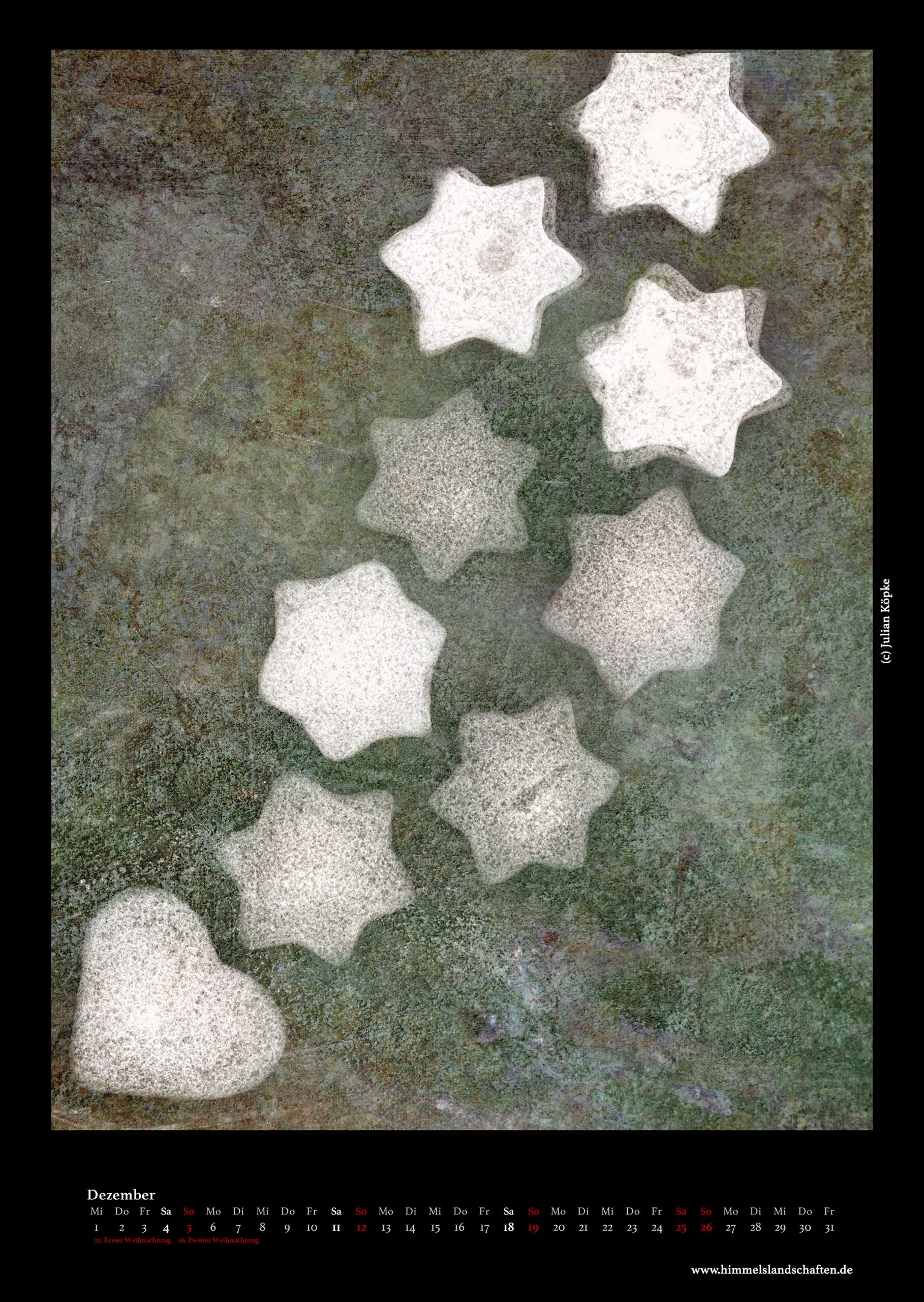
Calendar 2021
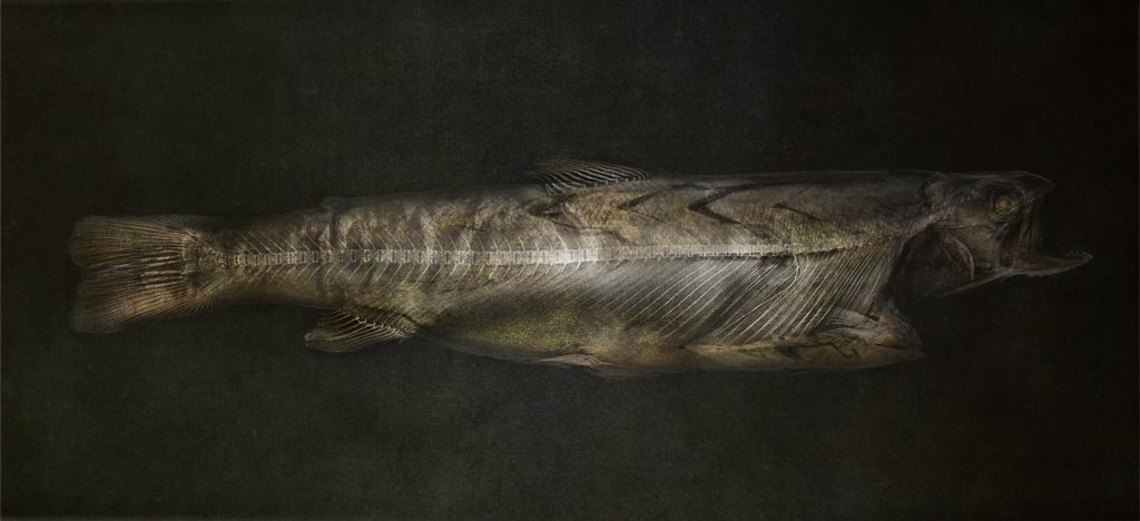
Trout X-ray fusion photo © Julian Köpke 
Black swan: pumpkins and fir cones X-ray photo © Julian Köpke 
-
New X-ray fusion photo compositions

Fusion X-ray photo of bananas © Julian Köpke 
Fruit bowl X-ray fusion photo © Julian Köpke 
X-ray fusion photo of lychees in a wooden bowl © Julian Köpke 
X-ray fusion photo of lychees and fruit in a wooden bowl © Julian Köpke 
Fruit X-ray fusion photo © Julian Köpke -
Snail shells X-ray fusion photos

X-ray fusion photo snail shells composition II © Julian Köpke 
X-ray fusion photo snail shells composition III © Julian Köpke 
X-ray fusion photo snail shells composition IV © Julian Köpke -
X-ray fusion photo of a sphere of snail shells

X-ray fusion photo of a sphere of snail shells © Julian Köpke 
Sphere of snail shells © Julian Köpke -
Calendar 2020

-
Spider conch X-ray fusion photo

Fusion image spider conch X-ray and photo seen from the bottom © Julian Köpke 
Fusion image spider conch (lambis lambis) X-ray and photo seen from above © Julian Köpke 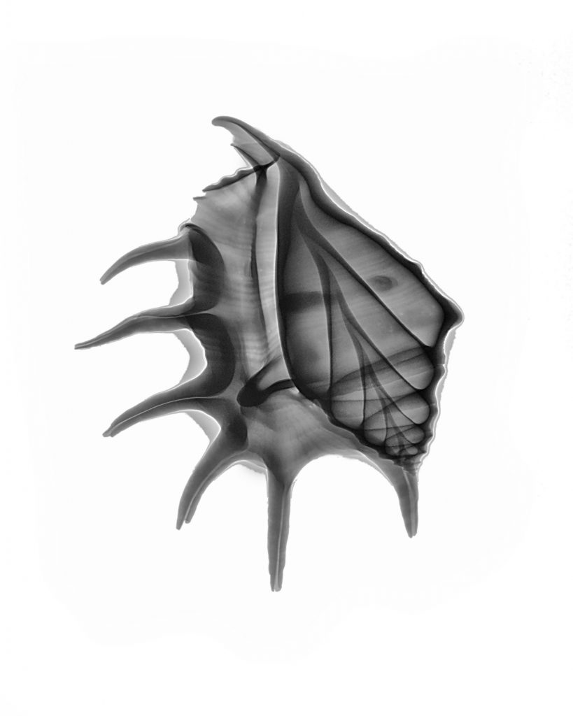
Spider conch (lambis lambis) © Julian Köpke 
Spider conch (lambis lambis) © Julian Köpke -
Orchid X-ray fusion photo

Orchid fusion X-ray photo © Julian Köpke -
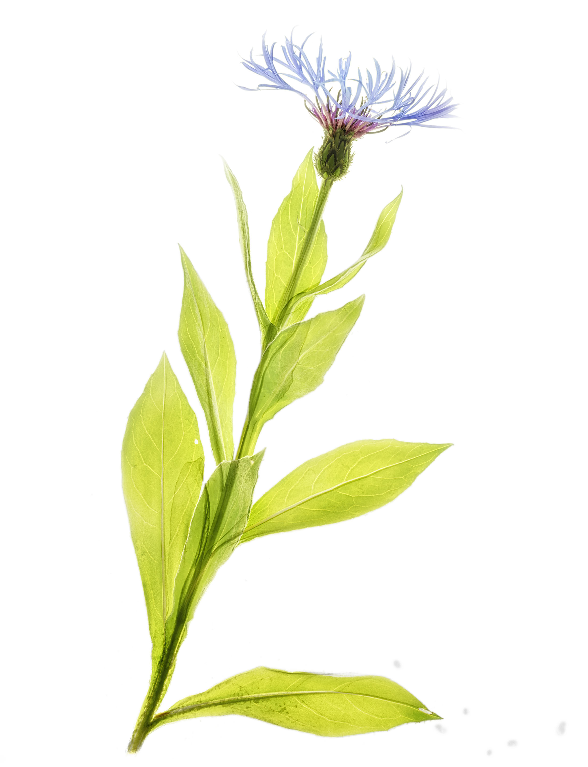
Spring and X-ray fusion photos

Cornflower X-ray fusion photo © Julian Köpke 
Blue aquilegia X-ray fusion photo © Julian Köpke -
Gradients and X-ray tubes

Gradient in an X-ray of 6 red calla lilies © Julian Köpke 
Gradient in an X-ray of 6 red calla lilies, inverted for creative reasons © Julian Köpke -
Effect of photon energy on X-ray images

Rose digital X-ray photo at 40kV © Julian Köpke 
Rose digital X-ray photo at 90kV © Julian Köpke 
Rose digital X-ray photo at 60kV © Julian Köpke 
Rose digital X-ray photo at 109kV © Julian Köpke


