• flowers,  Fusion imaging

    Purple Clematis

    Long lasting blossoms, turning up every year: my purple clematis in our garden.

    It was my third X-ray session with flowers this week. Third fusion imaging attempt. After blue cornflower and blue aquilegia now a purple clematis. Big data on my hard disk.

    Today we did it with mammography at 30 kV and 50 mAs. Lower noise ! Here is the positive representation of a single clematis:

    Clematis X-ray photo © Julian Köpke

    I processed the lightbox HighKey series with a mask. There was a shift of 2 or 3 pixels from the lightest to the darker images. So I processed everything a second time to compensate for the shift. The HDR image shows a cut stalk. Photoshop is made for this.

    Purple Clematis © Julian Köpke

    The stalk can be lengthened like in the preceding X-ray. The fusion image shows hidden leaves, the core of the blossom and stalks much better:

    Purple Clematis X-ray fusion photo © Julian Köpke

    The flower looks pretty fragile now, close to its natural appearance.

  • Fusion imaging,  Lightbox,  X-Ray

    Spring and X-ray fusion photos

    First flowers in spring show up. With much support from my colleagues I’m able to do some fusion images. We all would like to have another calendar.

    Preparing the lightbox, the X-ray machines, my camera and picking out the data is a bunch of hassle.

    My personal favorite is the blue cornflower. It looks like a print of an old botanic book:

    Cornflower X-ray fusion photo © Julian Köpke

    The next day I turned my attention to our white and blue Aquilegias. No chance to process the raw data yesterday. Eventually, there was a chance today, after quite a bit of tedious work at my desk:

    Blue aquilegia X-ray fusion photo © Julian Köpke
  • X-Ray

    Gradients and X-ray tubes

    Fusion imaging is a method full of surprise. My red calla lilies revealed an effect I had forgotten completely. There must be a gradient in every X-ray exposure.

    Preparing a fusion image composition with my 6 red calla lilies I found a troublesome gradient in the X-ray. 

    Gradient in an X-ray of 6 red calla lilies © Julian Köpke

    The cause for the gradient is a weakening of X-ray radiation at its origin in the X-ray tube. A closer look at the phenomenon can be found in my FAQ. This effect of variable recording of photons phycisists call „anode heel effect“.

    As part of my creative process I rotated the composition shown above by 180 degrees and exposed it a second time with the same parameters. Note that post-production as well was done equally for both X-ray exposures !

    Gradient in an X-ray of 6 red calla lilies, inverted for creative reasons © Julian Köpke
  • X-Ray

    Effect of photon energy on X-ray images

    I assume that everyone has had at some point the experience where less was more. Especially when dealing with computer based image postproduction. Software makes handy wonderful, or better: powerful, filters. Experienced artists know that only a pinch of something or homeopathy is a key to better results.

    The same holds true in X-ray production. A maximum of energy does not provide better images. Let’s look closer at this point.

    What is the influence of energy to X-ray images ?

    Higher energies in X-rays mean shorter wavelengths and a higher resolution. Therefore it might seem reasonable to increase the energy in our X-ray tubes always to the maximum to produce incredible images based on a maximum resolution.

    With four images below I show the influence of increased energy levels on X-ray images of a single rose. The applied energy levels are 40kV, 60kV, 90kV and 109kV. The steps of postproduction were the same in every image. Slight differences are owed to best contrast in each exposure.

    Surprisingly to the novice we get an increasing loss of contrast (or less available contrast) in each image with higher energies. This effect of loosing contrast can easily be seen in this series of four X-rays and is highest at 109kV.

    Rose digital X-ray photo at 40kV © Julian Köpke
    Rose digital X-ray photo at 90kV © Julian Köpke
    Rose digital X-ray photo at 60kV © Julian Köpke
    Rose digital X-ray photo at 109kV © Julian Köpke

    The explanation for less available contrast with higher energies is the following physical effect: the more photons have shorter wavelengths the more photons run unaffected through the object down onto the sensor. With all photons running through without any hindrance the sensor would show a homogenous gray value.

    Every structure looses contrast when turning to higher energies. The optimum for a structure is found by experience and varies significantly.

    In the medical field the applied energy strongly depends on the purpose of the examination and the structural demands to be diagnosed.

    The above demonstrated meaningless low contrast for our single rose at 109kV doesn’t hold true at all in radiology. Radiologists use frequently 125kV for a chest film to get reproducibly valuable contrast in most patients.

  • X-Ray

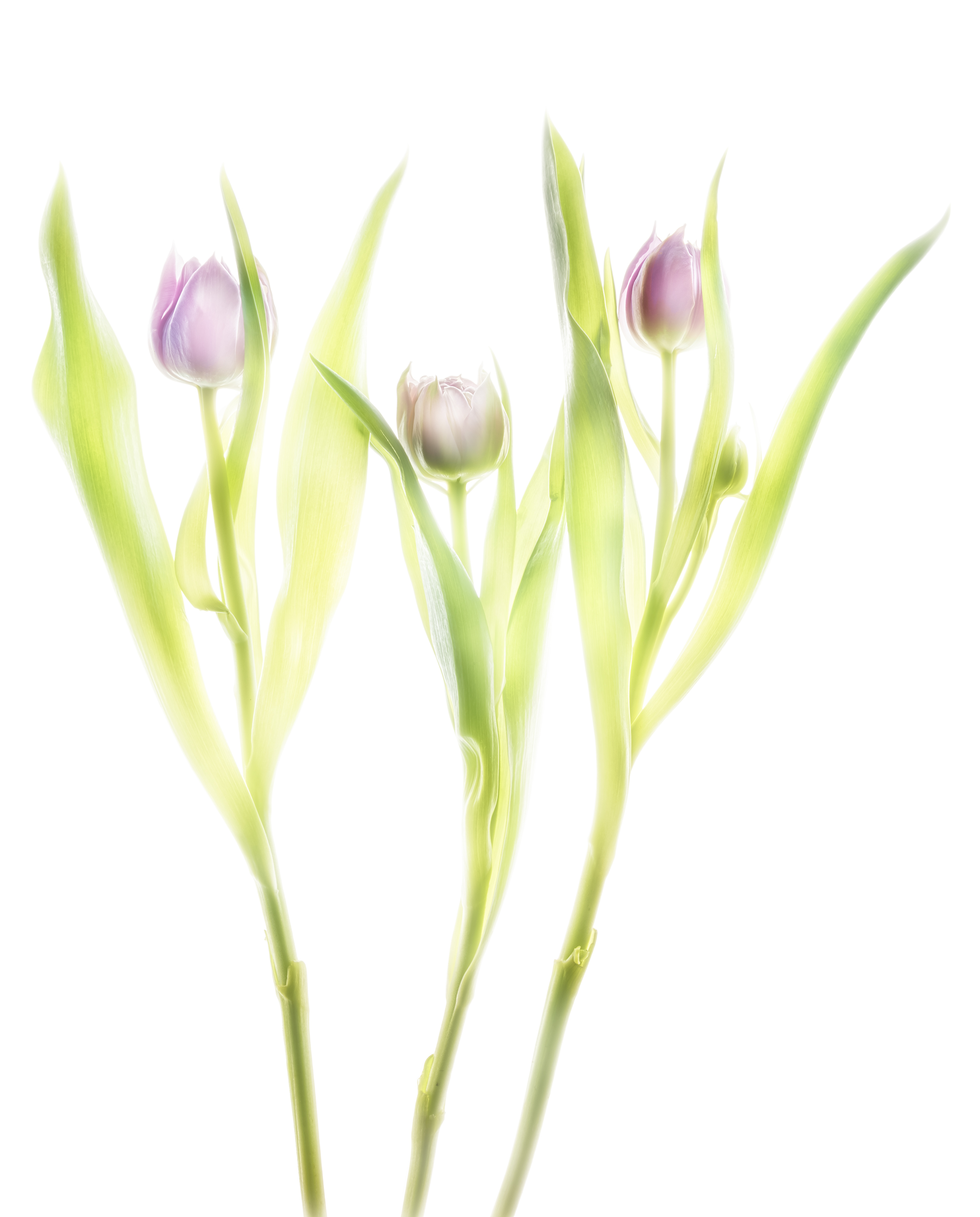
    Fusion X-ray of tulips

    Fusion imaging is beauty made of composite X-ray images and HDR images on a light box. The primary question is what energy fits best for flowers. To my experience 40 kV is often suitable. But: the proof of the pudding is in the eating.

    Mammography systems start e.g. from 20 kV and reach 39 kV. The sensor is up to 24cm x 30cm. Conventional systems start from 40 kV and reach 125 kV. The sensor is up to 43cm x 43cm what makes them more attractive to floral compositions.

    The higher resolution and the lower energies of a mammography will suit better for transparent objects. But the spatial limit of a composition (which is 24cm x 30cm) might put hard restrictions on the artist.

    Floral compositions have more creative space with a bigger sensor. But the X-ray tube starts with 40 kV and this might lead to overexposure of tender structures.

    Thus I performed today more than ten compositions to study this relation.

    After four exposure of three tulips I found this composition with four dense blossoms attractive to go further. The composition might somehow resemble to a sketch of three angels. The image is nice due to very soft edges of their „wings“, technically blown out portions in the image.  The inner structure of the nearly closed blossoms is well resolved. The stalks serve as „body“. There is no advantage with higher energies.

    X-ray three tulips © Julian Köpke

    The same composition was done immediately after the X-ray as a bracketing series on a lightbox. After returning I processed a manual HDR, the colors not to warm.

    Three purple tulips HDR photo © Julian Köpke

    The final fusion image is a composite of the preceding two images. Compared to the lightbox photo, the hidden stalks reappear naturally, the inner petals are outlined like a sketch.

    Three purple tulips fusion X-ray photo © Julian Köpke
  • Lightbox,  X-Ray

    X-ray fusion photo of a Nautilus

    Fusion imaging can be done retrospective. My split Nautilus shell on a light box rendered with manual HDR shows already a nice structure of the inner parts. 

    Nautilus shell manual HDR photo on a light box © Julian Köpke

    The X-ray obtained a couple of days earlier easily fits onto the HDR with not a big deal of processing.

    Nautilus X-Ray Energy Compressed © Julian Köpke

    The meaning of the fusion image may be different to the flowers. But it’s feasible to do it retrospectively.

    Nautilus shell fusion X-ray photo and manual HDR photo on a light box. © Julian Köpke
  • X-Ray

    Nautilus shell X-ray fusion photo of energy levels

    Different energies of X-ray radiation mean different transparency of an object. There is an example in my FAQ using a Nautilus shell.

    Instead of compressing images of different energies to a single image today I subtracted the 70 kV image of a Nautilus shell from the 40 kV image.

    The central parts of the Nautilus shell are more dense and show a significant higher difference. The core of the shell gets shiny. This is how it looks like:

    Energy difference X-ray photo of a Nautilus shell. The image is the difference of a 70kV and a 40 kV image. © Julian Köpke

    In positive X-ray representation you can compare the results. Left hand is the compressed image of 4 different energy levels, right hand the difference image.

    Nautilus X-Ray Energy Compressed © Julian Köpke
    Energy difference X-ray photo of a Nautilus shell. The fusion image is the difference of a 70kV and a 40 kV image in X-ray positive representation. © Julian Köpke
  • X-Ray

    Dahlias fusion X-ray HDR photo

    Long time ago my friend Harold and I did these X-rays in my practice. There was so much to do. Today was a chance to process the fusion images. Some details can be found in my FAQs.

    The manual HDR is already appealing to our eyes.

    Dahlias using manual HDR in visible light

    There is some charm in the X-ray image of the same composition. The hidden parts of the stalks can be clearly seen. 

    Five Dahlias X-ray photo © Julian Köpke

    The fusion image of this composition shows both color and hidden structures.

    Dahlias fusion digital X-ray with manual HDR photo in visible light

    Finished image with a background:

    Dahlias fusion digital X-ray and manual HDR photo with background © Julian Köpke
  • X-Ray

    Sunflower X-ray photos revisited

    How to show the sun in the middle of a sunflower ? For astronomers it is quite common to look at the sun in hydrogen alpha light, which is a pure red at 635nm. With artistic eyes, a red center might be overdone.

    So I tried two different representations, one in BW that is close to the natural look and feel of a sunflower and one with a light blue in the center as complementary color to the yellow petals.

    This sunflower is a fusion photo of X-ray, monochromatic Hα light of the sun converted to BW and a sunflower on a lightbox. © Julian Köpke
    This sunflower is a fusion photo of X-ray, monochromatic Hα light of the sun converted to blue and a sunflower on a lightbox. © Julian Köpke

    The surface structure of our sun can be seen like astronomers see it.

    There is no photo of the next digital X-ray image of a sunflower with its stalk and a leaf:

    Digital X-ray photo of a sunflower (inverted representation). © Julian Köpke
  • X-Ray

    Shells fusion X-ray photo

    Long time I dreamed of this fusion image of shells. Because already on a lightbox some of the shells are transparent and have nice colors. I like the shining through effect very much.

    The X-ray image is a compromise of structure and density resolution, depending on the maximum energy the mammography system is able to produce.

    Today I’m not at all in a stable state due to a recurrent infection. So I allowed me to do this image instead of hard working.

    Shells fusion X-ray photo © Julian Köpke

    It is the light inversion in Lab color mode that shows more of a X-ray look and feel. The colors are pretty close to the bright image.

    Shells fusion X-ray photo with inversion of the L-channel © Julian Köpke