-
X-ray photos of grapes and sunflowers

Grapes picture in Hologic calendar September 2019 © Julian Köpke 
Grapes - creative representation of an X-ray with Lab color © Julian Köpke 
Grape X-ray photo © Julian Köpke 

Grapes X-ray photo X-ray mammography photo © Julian Köpke 
Grapes composition II X-ray mammography photo © Julian Köpke 
I dedicate this X-ray of a bouquet of sunflowers to my colleague Dr. Arendt. © Julian Köpke 
Bouquet of Sunflowers X-ray photo © Julian Köpke -
Vegetables X-ray photography

Corncob X-ray photo © Julian Köpke 
Chickory (Belgian endive) and lettuce X-ray photo © Julian Köpke 
Chickory (Belgian endive) and lettuce mammography X-ray photo © Julian Köpke 
Onions mammography X-ray photo © Julian Köpke 
Onions mammography X-ray photo © Julian Köpke 
Stacked onions X-ray photo © Julian Köpke 
Fennel © Julian Köpke 
Sleeping beauty's choice © Julian Köpke 
An apple a day keeps the doctor away © Julian Köpke -
Purple Clematis

Clematis X-ray photo © Julian Köpke 
Purple Clematis © Julian Köpke 
Purple Clematis X-ray fusion photo © Julian Köpke -
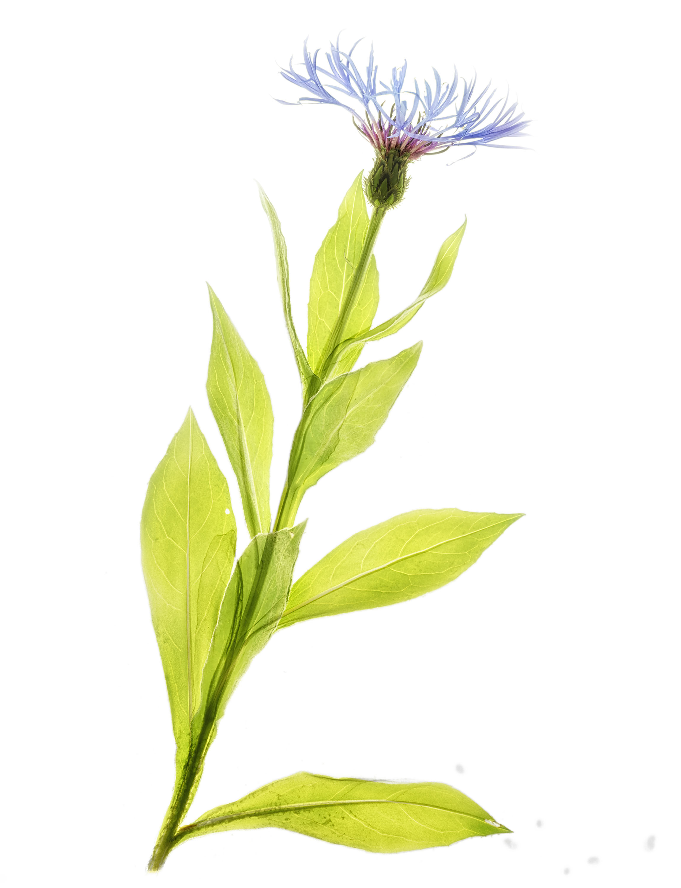
Spring and X-ray fusion photos

Cornflower X-ray fusion photo © Julian Köpke 
Blue aquilegia X-ray fusion photo © Julian Köpke -
X-ray images
-
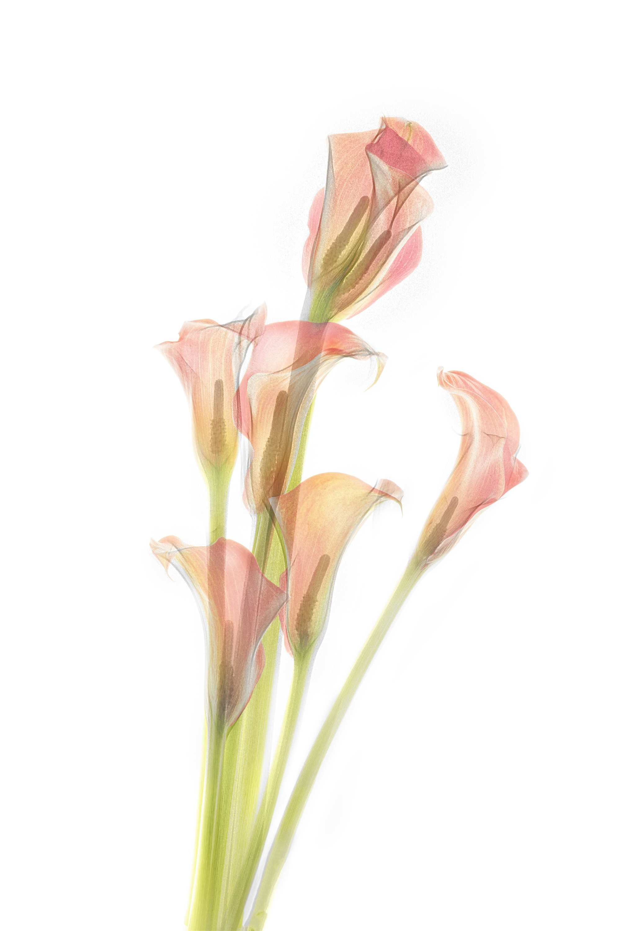
Red calla lilies

Fusion X-ray photo Calla lilies IV © Julian Köpke 
Fusion X-ray photo Calla lilies IV. Black background using Lab inversion. © Julian Köpke -
Red calla lilies

Calla lilies III © Julian Köpke 
Calla lilies with a twin blossom right hand side atop a twin stalk. © Julian Köpke -
X-ray exam of stone age tusk

Tip of a mammoth tusk (CT scan, VRT and Photoshop) © Julian Köpke -
X-ray fusion photo of a Nautilus

Nautilus shell manual HDR photo on a light box © Julian Köpke 
Nautilus X-Ray Energy Compressed © Julian Köpke 
Nautilus shell fusion X-ray photo and manual HDR photo on a light box. © Julian Köpke -
Tilted Nautilus X-ray photo

Tilted Nautilus digital X-ray photo. © Julian Köpke


